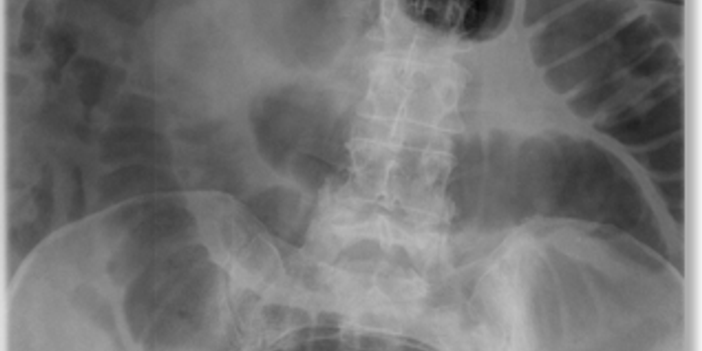
Small bowel obstruction is treated first with hospital-based resuscitation, bowel rest, intravenous fluids, electrolyte correction, pain relief, and selective nasogastric decompression. If the blockage does not resolve—or if there is strangulation, ischaemia, perforation, peritonitis, or a closed-loop process—urgent surgery may be required.
CT imaging and structured monitoring help clinicians decide whether an intestinal obstruction can be managed without an operation. Any person with persistent vomiting, severe abdominal pain, increasing distension, or inability to pass gas should seek emergency assessment.
Key takeaways
- Adhesions from previous surgery are the commonest cause in the UK, then herniae.
- Small bowel dilated beyond 3 cm, with valvulae conniventes crossing the full width of the lumen, is the plain-film finding.
- Drip and suck: nil by mouth, a nasogastric tube, and intravenous fluid and electrolyte replacement.
- CT identifies the level, the transition point, the cause and the complications.
- Strangulation or a closed loop means an operation, not a trial of conservative management.
What Is a Small Bowel Obstruction?
Small bowel obstruction is a partial or complete blockage that prevents food, fluid, gas, and intestinal contents from passing through the small intestine. The blockage may be mechanical or functional. Mechanical obstruction occurs when a physical barrier narrows or closes the bowel lumen. Ileus is a functional obstruction, where bowel movement stops or becomes severely reduced without a physical blockage. Common triggers for ileus include surgery, infection, electrolyte disturbance, and certain medicines.
Intestinal obstruction is a condition in which material cannot move normally through the intestines. The terms intestinal obstruction and bowel obstruction are often used interchangeably, but the location and underlying cause determine the safest management.
The symptoms bowel obstruction commonly include cramping, abdominal swelling, nausea, vomiting, and reduced bowel motions. A partial blockage may still allow some stool or gas to pass, while a complete blockage usually prevents both.
Common causes
In the UK, adhesions from previous abdominal surgery are the commonest cause, followed by herniae. Adhesions are bands of scar tissue that can kink or compress the bowel. Other causes include:
- Tumours or other intestinal malignancy
- Crohn’s disease causing inflammation and strictures
- Volvulus, where the bowel twists around its mesentery
- Food bolus or foreign material
- Congenital bands, particularly in younger patients
Adhesions account for approximately 65–75% of cases in developed countries. The main causes bowel obstruction include adhesions, hernias, tumours, twisting, inflammatory narrowing, and impacted material. The causes bowel obstructions differ by age, previous operations, cancer history, and the presence of inflammatory disease.
Scar tissue may develop after an abdominal operation, infection, or inflammation. This scarring can create a band that pulls, kinks, or compresses the intestine. A scar or internal band may cause recurrent episodes even years after the original operation.
Symptoms and examination findings
Symptoms of typical symptoms include colicky central abdominal pain, early vomiting, abdominal distension, constipation, and failure to pass flatus. Vomiting may occur earlier and more prominently than with large bowel obstruction.
Cramping often occurs in waves as the intestine attempts to push contents past the blocked area. Constant abdominal pain is more concerning because it can indicate ischaemia, perforation, or another complication.
Examination may reveal a dehydrated patient, tachycardia, abdominal distension, and visible peristalsis. Early in the illness, bowel sounds may be high-pitched or “tinkling”. Later, sounds can become quiet or absent. Palpate for a mass, previous surgical scars, and external herniae. A rectal examination may show an empty rectum.
Plain abdominal imaging may show central, dilated small bowel loops. The small bowel is typically dilated beyond 3 cm, with valvulae conniventes crossing the full width of the lumen. CT can identify the obstruction’s level, cause, and complications.
Why early recognition matters
Persistent obstruction increases pressure within the bowel. This can impair venous drainage, then arterial blood flow, causing strangulation and bowel ischaemia. Severe or constant pain, fever, tachycardia, peritonism, metabolic acidosis, or clinical deterioration suggests possible compromised bowel and requires urgent senior and surgical assessment.
Small bowel obstruction is a potentially life-threatening failure of intestinal transit, so identify its cause and look early for strangulation or ischaemia. > An intestinal obstruction should be treated as an emergency when pain becomes constant, the patient deteriorates, or imaging suggests compromised tissue.
How do you assess and stabilise the patient?
Suspected small bowel obstruction requires simultaneous resuscitation, diagnosis and early surgical involvement. Treat the patient, not only the abdominal X-ray.
Immediate assessment
- Begin with an ABCDE assessment, while checking for shock, respiratory compromise, sepsis and severe dehydration.
Give oxygen if hypoxic, attach monitoring, record observations, and obtain urgent senior help for instability.
- Take focused history about pain, vomiting, distension, constipation, previous operations, herniae, malignancy and inflammatory intestinal disease.
Adhesions from previous surgery are the commonest UK cause, followed by herniae; other causes include tumours, volvulus and food bolus.
- Examine the abdomen for distension, scars, tenderness, guarding, rigidity, masses, bowel sounds and signs of peritonitis.
Complete a careful groin, femoral, umbilical and incisional hernia examination, because a trapped hernia can rapidly strangulate bowel.
- Perform a digital rectal examination when clinically appropriate, documenting stool, blood, masses and whether the rectum is empty.
An empty rectum supports distal intestinal obstruction, but a normal examination does not exclude a serious blockage.
- Insert two large-bore intravenous cannulas, send blood tests, and begin isotonic crystalloid for hypovolaemia or circulatory compromise.
Request FBC, urea and electrolytes, creatinine, glucose, lactate, CRP, clotting, group and save, and crossmatch when surgery seems likely.
- Correct potassium, sodium, chloride and acid–base abnormalities carefully, replacing gastrointestinal losses while reassessing observations and urine output.
Insert a urinary catheter in an unwell patient, targeting at least 0.5 mL/kg/hour output.
- Keep the patient nil by mouth, prescribe suitable intravenous analgesia and antiemetics, and reassess pain after treatment.
Adequate analgesia does not prevent diagnosis; worsening pain despite treatment may indicate ischaemia or strangulation.
Decompression and escalation
- Use nasogastric decompression for persistent vomiting, marked distension, high-grade obstruction, aspiration risk or substantial intestinal fluid accumulation.
Record the tube position and output, provide suction as directed, and replace large ongoing losses with appropriate intravenous fluids and electrolytes.
- Request urgent senior surgical review for peritonism, shock, rising lactate, fever, tachycardia, continuous pain or suspected closed-loop obstruction.
These red flags suggest strangulation, ischaemia, perforation or sepsis and may require immediate laparotomy rather than observation.
- Arrange urgent imaging and operative planning when the patient deteriorates or conservative treatment fails.
CT usually identifies the obstruction’s level, transition point, cause and complications; plain films may show small bowel dilated beyond 3 cm, with valvulae conniventes crossing the full lumen.
The initial “drip and suck” approach means nil by mouth, nasogastric decompression, intravenous fluids and electrolyte replacement. However, unresolved obstruction or compromised bowel requires definitive surgical management.
Stabilise first, decompress selectively, examine for strangulation, and involve senior surgeons early when bowel viability is threatened.
How Is Small Bowel Obstruction Diagnosed?
A small bowel obstruction is a blockage that prevents intestinal contents and gas passing through the bowel. Diagnosis must confirm mechanical obstruction, identify its causes, and detect complications. Adhesions, herniae, tumours, volvulus, and inflammatory narrowing are common causes. History, examination, blood tests, and imaging all contribute, including in subacute or chronic presentations (clinical guidance on diagnosing subacute or chronic intestinal obstruction).
The direct answer is to request blood tests and cross-sectional imaging, usually a contrast CT of the abdomen and pelvis. Plain abdominal radiographs may support the diagnosis, but CT provides more information. It shows the level and cause of the obstruction and whether strangulation, ischaemia, perforation, or a closed-loop process is present.
Symptoms of types bowel obstruction include mechanical and functional obstruction, as well as partial, complete, simple, and closed-loop patterns. Another useful distinction is location: the small intestine differs from the large intestine in anatomy, diameter, symptoms, and common causes.
Small intestine large intestine comparisons matter clinically. A large bowel obstruction often produces more marked constipation and distension, whereas a blockage higher in the small intestine may cause earlier vomiting. However, symptoms overlap, so imaging is important.
Blood tests
Blood tests assess dehydration, inflammation, intestinal ischaemia, and readiness for surgery. Request:
- Full blood count, including haemoglobin and white cell count
- Urea, creatinine, and electrolytes
- C-reactive protein or another inflammatory marker
- Venous or arterial blood gas, including lactate
- Group-and-save testing
Raised white cells, inflammatory markers, or lactate may suggest strangulation or bowel ischaemia. However, normal results do not exclude serious disease. Renal function and electrolytes guide intravenous fluid replacement and assess the safety of contrast imaging. Group-and-save prepares for possible urgent surgery or bowel resection.
Imaging tests
An erect chest radiograph may show free air beneath the diaphragm. This suggests pneumoperitoneum from perforation and requires urgent surgical assessment. An abdominal radiograph can show central, dilated small-bowel loops. Small bowel is typically dilated beyond 3 cm, with valvulae conniventes crossing the full bowel width.
Ultrasound can identify dilated, fluid-filled loops and abnormal bowel movement. It may help when avoiding radiation, such as in pregnancy. However, bowel gas, obesity, and operator experience limit its accuracy. Plain films also cannot reliably identify the cause or bowel viability.
Contrast CT abdomen and pelvis is usually the most useful investigation in adults. It can demonstrate dilated proximal intestine, collapsed distal bowel, and a transition point. It may also identify adhesions indirectly, herniae, tumours, volvulus, or inflammatory causes. CT findings of reduced enhancement, mesenteric oedema, free fluid, pneumatosis, or portal venous gas raise concern for ischaemia.
A closed-loop obstruction occurs when one bowel segment is blocked at two points. CT may show C-shaped or U-shaped loops, twisted mesenteric vessels, and free fluid. Pneumoperitoneum suggests perforation rather than uncomplicated obstruction.
These findings guide treatment. Stable patients without ischaemia, perforation, or peritonitis may receive conservative “drip and suck” management. Free air, a closed loop, worsening pain, acidosis, rising lactate, or suspected strangulation requires urgent surgery.
CT confirms the obstruction, explains its cause, and identifies the complications that determine whether conservative treatment or surgery is safest.
When can you manage it without surgery?
Stable patients without peritonitis, ischaemia, strangulation or perforation may receive a monitored trial of non-operative care. This involves bowel rest, intravenous resuscitation, electrolyte correction and nasogastric decompression, with early surgery if the patient deteriorates or fails to improve.
When conservative treatment is appropriate
Non-operative management is most suitable for a stable patient with a likely adhesive small bowel obstruction. Adhesions commonly follow previous abdominal surgery. A hernia, tumour or volvulus may require earlier surgical treatment.
Patients should not undergo prolonged conservative care when there are signs of bowel compromise. Peritonism, continuous severe pain, fever, tachycardia, shock, rising lactate, metabolic acidosis or CT evidence of ischaemia require urgent senior surgical review.
Patients without these warning signs can usually receive an initial non-operative trial. Complete obstruction has a higher failure rate than partial obstruction, so the threshold for escalation should remain low.
The principles of “drip and suck”
Drip and suck means nil by mouth, intravenous fluids, electrolyte replacement and nasogastric decompression. Bowel rest reduces further intestinal distension. Intravenous crystalloid replaces losses from vomiting and third-space fluid shifts.
Potassium, sodium, chloride and magnesium abnormalities should be corrected. Blood tests should also assess renal function, glucose, full blood count, C-reactive protein and acid-base status.
A nasogastric tube drains gastric contents and reduces vomiting, aspiration risk and proximal bowel distension. Record its output, along with urine output, stool, flatus and total fluid balance. A urinary catheter may help monitor resuscitation in an unwell patient.
Contrast studies and monitoring
In selected adhesive obstruction, a water-soluble contrast study can help predict whether the blockage will resolve. Contrast reaching the colon on follow-up imaging supports successful non-operative treatment. Failure to progress suggests persistent obstruction and may support operative management.
Review the patient regularly, rather than relying on one normal examination. Monitor pain pattern, abdominal tenderness, guarding, distension, bowel sounds, hernias, vomiting and passage of flatus. Repeat observations and measure fluid balance accurately.
Repeat biochemical tests to identify dehydration, renal injury, electrolyte loss, rising inflammatory markers or lactate. New tachycardia, worsening pain, peritonism, fever, oliguria or acidosis should prompt immediate reassessment.
If symptoms, abdominal signs, obstruction on imaging or biochemical abnormalities fail to improve during the agreed observation period, discuss surgery urgently. Deterioration at any stage overrides the conservative trial. The aims are to prevent ischaemia, necrosis and perforation, while treating the cause of the small bowel obstruction.
Water-soluble contrast protocols, CT findings, and repeated clinical examinations are used together rather than as isolated tests. The decision that bowel obstruction is treated without an operation should therefore be reviewed as the patient’s condition changes.
When should non-operative treatment stop? Stop and escalate when the patient deteriorates, develops signs of bowel compromise or fails to show clinical and biochemical improvement.
Conservative care is appropriate only for carefully selected patients, with active monitoring and early surgery when obstruction does not resolve.
When does it need an operation?
What are the indications for surgery?
Surgery is needed when an intestinal obstruction threatens the bowel or the patient’s life. Peritonitis, perforation, strangulation, or suspected ischaemia are emergency indications. These findings suggest compromised blood supply, leakage, or infection within the abdomen; clinical reports of small-bowel perforation also emphasise the need for careful assessment and timely escalation (case discussion of small-bowel perforation and surgical assessment).
Other urgent indications include:
- A closed-loop obstruction, where one bowel segment is blocked at two points
- A complete obstruction with persistent vomiting and no passage of flatus
- A tender, irreducible hernia causing the obstruction
- Worsening pain, tachycardia, fever, acidosis, or rising lactate
- Deterioration despite intravenous fluids, nasogastric drainage, and electrolyte replacement
- Persistent or recurrent obstruction despite appropriate non-operative treatment
Adhesions from previous surgery are the commonest cause in the UK, followed by herniae. Other causes include tumours, strictures, volvulus, inflammatory disease, and an obstructing food bolus. A recurring obstruction, especially with a palpable mass, may justify earlier resection after adequate medical treatment.
An intestinal obstruction caused by a tumour requires particular attention to bowel cancer and colon cancer. Malignancy can narrow the passage directly or produce scarring and adhesions. The causes bowel obstruction assessment should therefore include weight loss, anaemia, previous cancer, altered bowel motions, and a palpable mass.
Why does timing matter?
A closed-loop obstruction can rapidly increase pressure within the intestine. This reduces venous drainage, then arterial flow, causing bowel ischaemia and necrosis. Perforation may follow, leading to peritonitis, sepsis, and death.
Partial obstruction may settle with careful observation and “drip and suck.” However, complete obstruction, suspected strangulation, or clinical deterioration usually requires immediate surgery. A patient should not undergo prolonged conservative treatment when examination or blood results suggest a threatened bowel.
How is the operation performed?
An emergency laparotomy provides rapid access through a midline abdominal incision. The surgeon identifies the transition point, examines the entire intestine, and releases the blockage. Operative options include:
- Adhesiolysis: dividing bands that kink or trap the bowel.
- Hernia repair: reducing the intestine and repairing the abdominal wall defect.
- Resection: removing non-viable or perforated bowel, then joining healthy ends where safe.
- Cause-specific treatment: managing a tumour, volvulus, stricture, or foreign body.
Laparoscopic surgery may reduce wound complications and recovery time in carefully selected, stable patients. It is less suitable with severe distension, widespread adhesions, peritonitis, haemodynamic instability, or suspected necrosis. Conversion to open surgery may be necessary.
Before theatre, continue nil by mouth, nasogastric decompression, intravenous fluids, and electrolyte correction. Insert a urinary catheter when close urine-output monitoring is needed. Give analgesia, antibiotics for suspected perforation or ischaemia, venous thromboembolism prophylaxis, and crossmatched blood when significant resection is possible.
Consent should cover bleeding, infection, enterotomy, stoma formation, anastomotic leak, further resection, intensive care, and death. Postoperative complications include ileus, recurrent obstruction, wound infection, pneumonia, acute kidney injury, sepsis, and anastomotic leak.
Symptoms of complications bowel obstruction include dehydration, kidney injury, strangulation, tissue death, perforation, peritonitis, sepsis, and recurrent blockage. A further complication can occur when delayed treatment allows a previously viable section of intestine to become necrotic.
What is the key MRCS answer? Operate for peritonitis, ischaemia, perforation, strangulation, closed-loop obstruction, or failure of resuscitation. Treat the cause, but operate promptly when obstruction threatens bowel viability or the patient’s stability.
What decisions will the examiner probe?
Small bowel obstruction is a mechanical blockage that prevents intestinal contents and gas passing through the small intestine.
Compare the management options
Treatment depends on the patient’s physiology, obstruction severity, and likely cause. Adhesions from previous surgery are the commonest UK cause, followed by herniae.
This “drip and suck” approach requires active reassessment. Record observations, abdominal findings, nasogastric losses, fluid balance, lactate, and inflammatory markers. CT findings and clinical deterioration should guide escalation. Malignant causes often carry a poor prognosis and require individualised planning, including consideration of palliative bypass approaches reported for patients with advanced cancer (a recent report of endoscopic bypass for terminal cancer-related obstruction).
The types bowel obstruction seen in examinations include adhesive, hernia-related, malignant, inflammatory, volvulus-related, and functional patterns. Intussusception is another possible mechanism, particularly in children, although an adult case may have a lead point such as a polyp or tumour.
A pseudo-obstruction produces symptoms resembling a blockage without a physical barrier. It can affect the small intestine or colon and may occur with severe illness, medications, metabolic abnormalities, or neurological disease. Distinguishing it from mechanical disease prevents inappropriate delay or unnecessary intervention.
Common MRCS Part B examination traps
Do not label every distended abdomen as mechanical obstruction. Ileus causes reduced intestinal movement, often after surgery, infection, electrolyte disturbance, or medication. It usually causes diffuse bowel dilation and quieter bowel sounds. Mechanical obstruction more often causes colicky pain, vomiting, and a transition point.
Do not delay surgery in suspected strangulation. Severe continuous pain, peritonism, fever, tachycardia, metabolic acidosis, raised lactate, or CT evidence of compromised blood supply requires urgent senior and surgical review. Early recognition of ischaemia and strangulation is central to safe management.
Practise the OSCE decision
Use interactive scenarios on Mrcspartbquestions to rehearse:
- Examination: assess an acutely unwell patient, identify shock, inspect for herniae, and perform an abdominal examination.
- Communication: explain nasogastric decompression, contrast imaging, possible surgery, risks, and consent.
- Surgical skills: demonstrate safe fluid prescribing, nasogastric tube care, catheterisation, and escalation.
MRCS Part B’s applied surgical science and critical care station awards 12 marks for knowledge and application, four for clinical and technical skill, and four for professionalism. Mrcspartbquestions provides realistic scenarios, explanations, mark schemes, and progress tracking to test applied understanding.
What is the key treatment principle? Stabilise first, identify complications early, and operate promptly when strangulation is suspected. In MRCS Part B, treat the patient’s physiology first, distinguish ileus from mechanical obstruction, and never delay surgery when strangulation is possible.
How is this topic marked in MRCS Part B?
MRCS Part B marks an applied surgical science and critical care station out of 20: 12 marks for clinical knowledge and its application, 4 for clinical and technical skill and 4 for professionalism. Three of the seventeen examined stations fall in this area.