Compartment syndrome requires urgent medical assessment when severe, escalating limb pain follows trauma, a fracture, surgery, bleeding, or compression. Early warning signs include pain out of proportion, pain with passive stretch, a tense compartment, numbness, and weakness; do not wait for a missing pulse before seeking emergency care.
Key takeaways
- Pain out of proportion to the injury, and pain on passive stretch of the compartment, are the earliest signs.
- Pulselessness is a late and unreliable sign: a normal pulse does not exclude compartment syndrome.
- The diagnosis is clinical; pressure monitoring supports it in the unconscious or unreliable patient.
- A delta pressure of 30 mmHg or less, diastolic minus compartment pressure, is the usual threshold to act on.
- Treatment is urgent fasciotomy, with wound review and closure once swelling settles.
What Is Compartment Syndrome?
Compartment syndrome is a rise in pressure inside a closed muscle compartment that reduces blood flow and damages muscle and nerve tissue. A compartment contains muscle, small blood vessels, and nerves, enclosed by tough, non-expanding fascia. As bleeding or swelling increases, the fascia cannot stretch enough to accommodate the extra volume.
Compartment syndrome pathophysiology is the cycle of increased compartment volume, impaired venous drainage, rising pressure, and reduced capillary perfusion. This pathophysiology explains why tissue can become ischaemic before a major artery is completely blocked.
Venous drainage becomes restricted first. Pressure then rises further, reducing the arteriovenous pressure gradient and capillary perfusion. Without oxygen, nerves and muscles become ischaemic. Acute compartment syndrome can cause permanent functional loss within hours and requires urgent surgical assessment.
Fascia is tough connective tissue that surrounds muscles and limits expansion. When bleeding or fluid accumulation occurs inside that space, pressures can rise faster than circulation can adapt.
The relevant anatomy differs by body region. The lower leg has four major compartments, while the forearm has tightly arranged flexor and extensor spaces. Understanding this anatomy helps clinicians identify which muscles, nerves, and vessels may be at risk.
Acute compartment syndrome most often follows a fracture, especially a tibial shaft fracture. Other causes include crush injury, reperfusion after ischaemia, bleeding disorders or anticoagulation, tight casts, and excessive swelling after exercise or trauma. A normal distal pulse does not exclude the condition. Pulselessness is a late and unreliable sign.
Compartment syndrome aetiology includes trauma, fractures, vascular obstruction, bleeding, reperfusion, burns, prolonged compression, and iatrogenic causes such as restrictive casts or intravenous fluid leakage. The same aetiology can affect a leg compartment, forearm, thigh, hand, or foot.
Emergency departments commonly combine repeated clinical examinations with pressure measurement, electronic records, and point-of-care documentation. However, no device replaces clinical judgement when the anatomy, mechanism, and examination strongly suggest an emergency.
Chronic exertional compartment syndrome develops when exercise repeatedly raises pressure in a compartment. Symptoms of chronic exertional compartment syndrome generally appear at a similar exercise intensity and improve with rest. The symptoms of chronic exertional compartment syndrome differ from the continuing pain and progressive neurological changes of an acute injury.
Is chronic exertional compartment syndrome an emergency? Usually not. Chronic exertional compartment syndrome needs clinical review, but acute compartment syndrome needs immediate assessment. Diagnosis is primarily clinical, supported by compartment pressure measurement when the examination is uncertain.
What is the key exam warning? Pain out of proportion to the injury and pain on passive stretch are early signs. Urgent fasciotomy is the definitive treatment for acute compartment syndrome.
Escalating pain and pain on passive stretch demand urgent assessment, even when pulses remain present. > Clinical insight: A palpable pulse confirms that some arterial flow remains; it does not prove that capillary blood flow inside the compartment is adequate.
What are the early signs of compartment syndrome?
Acute compartment syndrome is a limb-threatening emergency caused by rising pressure within a closed muscle compartment. Early recognition protects muscle and nerve function.
Compartment syndrome signs symptoms usually begin with disproportionate pain and may progress to pain on passive stretch, tense tissue, altered sensation, and weakness. The acute compartment syndrome symptoms pattern is progressive rather than reliably relieved by rest or ordinary analgesia.
The earliest warning symptoms
- Pain out of proportion to the injury is a key early warning sign, especially when ordinary analgesia no longer provides relief.
- Pain on passive stretch is an early clinical finding; stretch the muscles supplied by the suspected compartment gently and compare sides.
- A tense, swollen, or “wood-like” compartment should raise immediate concern, even when the skin colour and pulses appear normal.
- New paraesthesia, such as tingling or numbness, suggests nerve compromise and requires urgent reassessment rather than routine observation.
- Increasing weakness or pain during active movement indicates worsening muscle and nerve dysfunction within the affected compartment.
- A rising requirement for opioid analgesia can signal progression, particularly when pain remains severe despite appropriate treatment.
The condition may develop immediately or emerge up to 48 hours after the initial injury. Do not rely on one isolated finding. Serial examinations are essential, especially after fractures, crush injuries, vascular injury, reperfusion, burns, or tight casts and dressings.
Symptoms include severe pain, firm tissue, numbness, tingling, reduced movement, and increasing difficulty controlling the affected limb. These symptoms can be subtle in children, sedated patients, or people receiving strong analgesia.
The phrase symptoms compartment syndrome is often used in online searches, but the practical question is whether findings are worsening. Progressive pain, pain swelling, and neurological change require immediate action required by an emergency team.
Practical examination points
For the leg, inspect for increasing swelling and compare both sides. Palpate the anterior, lateral, superficial posterior, and deep posterior compartments for firmness. Assess passive ankle and toe movements. Passive toe flexion stretches the extensor compartments, while passive toe extension stretches the flexor compartments. Test sensation over the first web space and the dorsum and sole of the foot.
For the forearm, assess the volar and dorsal compartments. Pain on passive finger or wrist extension may indicate flexor compartment involvement. Check median, ulnar, and radial nerve sensation and motor function. Examine the hand for swelling, weakness, and altered sensation.
Also examine the thigh, buttock, hand, foot, and upper arm when injury or swelling affects these regions. Chronic exertional compartment syndrome usually causes exercise-related symptoms that settle at rest, unlike this acute emergency.
Pallor, paralysis, and pulselessness are late, unreliable signs. A normal pulse does not exclude the condition, so never wait for these findings before seeking senior orthopaedic review and urgent decompression.
Severe disproportionate pain, pain on passive stretch, and a tense compartment demand urgent action before pulses disappear. Compartment syndrome signs can change over minutes or hours, so a single reassuring examination is not always enough. Repeat documentation should include limb temperature, pulses, capillary refill, sensation, motor function, tissue firmness, and the patient’s response to analgesia.
What causes compartment syndrome?
Acute compartment syndrome occurs when pressure within a closed muscle compartment reduces capillary blood flow. Suspect it after trauma, surgery, vascular injury, or prolonged compression. The condition can develop even after apparently minor injuries.
Acute compartment syndrome happens suddenly or progresses rapidly when bleeding and oedema overwhelm the available space. This is different from chronic compartment syndrome, which generally follows a predictable activity pattern and improves after rest.
When should you suspect it?
The highest-risk traumatic situations include tibial fractures, forearm fractures, crush injuries, and penetrating trauma. High-energy trauma and vascular injury increase concern. A review found vascular injury had a pooled odds ratio of 9.05, while high-energy trauma had an odds ratio of 3.10.
Non-traumatic cases can follow anticoagulation-related bleeding, reperfusion, burns, prolonged pressure, or rhabdomyolysis. Tight plaster casts, circumferential dressings, splints, and external devices can compress the limb. They may worsen swelling by limiting the space available to the compartment.
The pathophysiology after a tibial fracture often involves bleeding from cancellous bone, muscle damage, and venous obstruction. As pressures rise, tissue oxygenation falls even if the tibial artery and distal pulse remain detectable.
Children, sedated patients, and those with reduced consciousness may not report the typical symptoms. Examine them repeatedly and compare both limbs. Pressure measurement can support the diagnosis when examination is unreliable, but a normal pulse does not exclude the condition.
Do not confuse this acute emergency with chronic exertional compartment syndrome, which usually causes exercise-related, recurring symptoms. In an MRCS scenario, unexplained escalating pain after injury demands urgent assessment and senior surgical review.
Which body areas are most vulnerable?
The anterior leg compartment is frequently discussed because it contains the tibialis anterior, extensor hallucis longus, extensor digitorum longus, and deep peroneal nerve. Anterior leg compartment syndrome may cause pain with passive plantarflexion and altered sensation in the first web space.
A leg compartment syndrome can involve the anterior, lateral, superficial posterior, or deep posterior space. The lower leg is particularly vulnerable after a tibial fracture because the fascia provides limited room for expansion.
A tibial fracture may be closed or open, displaced or nondisplaced, and still produce dangerous pressure. Clinicians should assess the tibial shaft, ankle, knee, and surrounding tissue rather than relying only on the appearance of the fracture.
The forearm has another important pattern. A forearm fracture can increase pressures in the volar compartment, while elbow trauma, vascular bleeding, or a tight dressing may threaten the flexor muscles, median nerve, and ulnar nerve.
Tibial anatomy, forearm anatomy, and compartment anatomy determine which passive movement produces pain. This is why a complete examination should identify the suspected space instead of recording only “limb pain.”
How Is the Diagnosis Confirmed?
Acute compartment syndrome is primarily a clinical diagnosis, and urgent treatment must not wait for investigations. Compartment pressure measurement supports the diagnosis when findings are unclear or the patient cannot report symptoms.
Clinicians diagnose compartment syndrome by combining mechanism, examination, progression, and, when necessary, measured compartment pressures. To diagnose compartment syndrome safely, the workup must account for the patient’s age, blood pressure, sedation, fracture pattern, and ability to communicate.
Clinical diagnosis comes first
Acute compartment syndrome is diagnosed from the history and examination. Severe pain out of proportion to the injury and pain on passive stretch are early warning signs. Tense swelling, altered sensation, and worsening neurological findings increase concern.
A normal distal pulse does not exclude the condition. Pulselessness is a late and unreliable sign. If clinical suspicion is high, urgent surgical review and fasciotomy should proceed without waiting for pressure testing or imaging.
The first diagnostic workup should record the mechanism, timing, fracture or trauma pattern, analgesic requirements, limb examination, and serial changes. Electronic observation systems such as Epic and Cerner can help display trends, but they cannot independently diagnose the syndrome.
Measuring compartment pressure
Pressure measurement can support the diagnosis when clinical findings are equivocal. It is particularly useful in patients with reduced consciousness, sedation, severe head injury, polytrauma, or distracting injuries. It also helps assess patients who cannot communicate their symptoms reliably.
A needle or catheter measures the pressure inside each relevant compartment. The result should be interpreted alongside the clinical picture, rather than in isolation. Measurements should be taken near the injured area and repeated if symptoms or examination findings change.
Delta pressure is the diastolic blood pressure minus the compartment pressure. A delta pressure of 30 mmHg or less is commonly considered concerning. StatPearls describes inadequate perfusion once the intracompartmental pressure comes within 10 to 30 mmHg of the diastolic blood pressure.
A single normal pressure reading does not reliably exclude evolving disease. Technical error, incorrect catheter position, delayed progression, or measuring the wrong compartment can produce false reassurance. Pressure monitoring should never delay decompression when the clinical diagnosis is clear.
Measured pressures are most useful when pressures are repeated across the relevant compartments and interpreted against systemic blood pressure. A low diastolic pressure can make a borderline compartment pressure more dangerous because the delta pressure is reduced.
What other tests show
Vascular examination and Doppler ultrasound assess arterial flow, but preserved flow does not rule out raised pressure. Creatine kinase may rise because of muscle injury. Urea, electrolytes, and creatinine help detect renal complications, including rhabdomyolysis-related acute kidney injury.
X-rays can identify fractures, while CT or MRI may show an underlying injury or alternative diagnosis. These investigations do not exclude raised pressure within a closed compartment. Chronic exertional compartment syndrome is assessed differently, using exercise-related symptoms and specialist pressure testing.
The key MRCS answer is: diagnose clinically, use pressure monitoring when examination is unreliable, and never delay urgent fasciotomy for a test. The diagnosis of chronic compartment syndrome uses a different workup. Clinicians may record baseline pressures, supervise exercise, reproduce the patient’s usual symptoms, and repeat pressures after exertion. This workup should also consider stress fracture, tendinopathy, nerve entrapment, vascular disease, and lumbar radiculopathy.
How is compartment syndrome treated?
Acute compartment syndrome is a surgical emergency requiring urgent decompression. Do not wait for pulselessness, paralysis, or pressure testing when the clinical findings are convincing.
Compartment syndrome treatments are selected according to urgency, but acute disease requires removal of external compression, resuscitation, surgical review, and fasciotomy. Conservative treatments cannot reliably relieve pressure once tissue perfusion is threatened.
In an MRCS Part B station, state your actions clearly:
- Call the senior orthopaedic or plastic surgeon immediately.
- Remove or split tight casts, dressings, splints, and bandages.
- Keep the limb at heart level, not elevated above the heart.
- Optimise circulation with oxygen, intravenous access, fluids, and treatment of hypotension.
- Repeat and document neurovascular observations.
- Prepare the patient for emergency theatre.
A normal distal pulse does not exclude this condition. Pulses are often preserved until late vascular compromise develops. Immediate action required means escalating to the senior surgical team, removing constriction, monitoring the patient, and preparing for emergency surgery. Do not elevate the limb excessively because this can reduce the arterial pressure gradient across an already compromised compartment.
Why is fasciotomy urgent?
Rising pressure within a closed compartment reduces capillary blood flow. Muscle and nerve ischaemia then progress to necrosis. The ideal target is decompression within six hours of injury, where possible. Delayed treatment increases the risk of permanent neurological deficit, contracture, infection, and amputation.
Fasciotomy is urgent because it opens the fascia and helps relieve pressure before muscle and nerve tissue become irreversibly ischaemic. The pathophysiology becomes progressively worse when venous obstruction, tissue oedema, and arterial inflow reduction reinforce one another.
How is definitive treatment performed?
The definitive treatment is urgent fasciotomy. The surgeon must release all affected compartments, not only the compartment with the most obvious symptoms. The diagnosis remains clinical, although pressure measurements can support decision-making in an unconscious, sedated, or polytrauma patient.
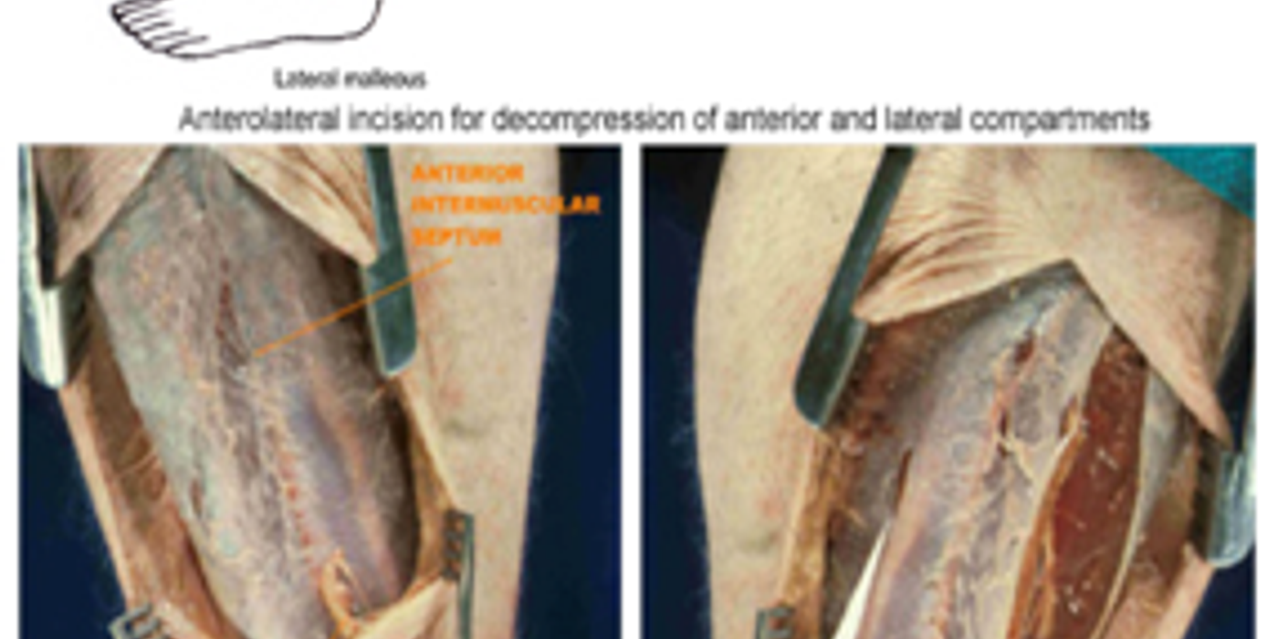
For a lower-leg injury, a standard double-incision approach releases:
- The anterior and lateral compartments through the lateral incision.
- The superficial and deep posterior compartments through the medial incision.
The surgeon must protect the superficial peroneal nerve laterally and the long saphenous vein medially. Intraoperative findings include tense muscle, reduced contractility, and poor bleeding. Non-viable muscle requires debridement.
Tibial fractures frequently require careful release of all four lower-leg compartments. The anterior compartment, lateral compartment, superficial posterior compartment, and deep posterior compartment must be assessed because tissue damage can extend beyond the visibly injured area.
What associated care is required?
Treat the underlying cause at the same time. Stabilise fractures with suitable splintage, external fixation, or definitive fixation when safe. Control ongoing haemorrhage and correct shock with blood products or intravenous fluids.
Monitor for rhabdomyolysis and acute kidney injury. Check creatine kinase, potassium, bicarbonate, creatinine, and urine output. Dark urine may indicate myoglobinuria. Hyperkalaemia, acidosis, and falling urine output require urgent senior and renal input.
Symptoms of compartment syndrome complications include muscle necrosis, nerve injury, infection, renal failure, contracture, and limb loss. These complications become more likely when treatment is delayed after trauma, fracture, or prolonged compression.
How are fasciotomy wounds managed?
Fasciotomy wounds are usually left open initially because swelling persists. Apply a non-adherent dressing, sterile gauze, and a light bandage. Plan a formal wound review and closure, often after 48–72 hours, once swelling and muscle viability have improved.
Delayed primary closure may be possible. Large defects may require negative-pressure therapy, skin grafting, or flap coverage. Early closure under tension risks recurrent pressure and tissue loss.
Symptoms of potential complications include infection, bleeding, muscle necrosis, nerve damage, joint contracture, chronic pain, and amputation. Chronic exertional compartment syndrome follows a different, planned pathway and does not usually require emergency fasciotomy.
Remove external pressure, restore perfusion, call senior help, and arrange urgent fasciotomy of every affected compartment.
How does chronic exertional compartment syndrome differ?
Chronic exertional compartment syndrome is a pressure-related condition that repeatedly develops during exercise and settles with rest. It differs sharply from acute compartment syndrome, which can rapidly threaten muscle and nerve function.
Chronic compartment syndrome is commonly used to describe exercise-related pressure elevation, although the more precise term is chronic exertional compartment syndrome. Chronic compartment syndrome usually has a reproducible threshold, unlike acute disease after trauma.
Chronic compartment syndrome can affect runners, military recruits, cyclists, and athletes who repeatedly load the lower leg. A gradual increase in training volume, footwear changes, or altered running mechanics may precede the presentation.
Recognising the clinical difference
Chronic exertional compartment syndrome usually follows a consistent pattern. A patient may report calf tightness or cramping after running for ten minutes. Paraesthesia may affect a predictable nerve distribution. Symptoms commonly resolve within minutes of stopping, although they return when the same activity restarts.
In contrast, acute compartment syndrome causes escalating pain, swelling, and neurological compromise. Pain may be out of proportion to the injury and worsen with passive stretch. These symptoms require urgent assessment. Do not wait for absent pulses, because vascular compromise is a late sign.
Chronic compartment syndrome does not usually follow a single fracture or crush event. Persistent symptoms at rest, night pain, marked tissue tension, or worsening neurological loss should prompt reassessment for acute disease, fracture, nerve compression, or another diagnosis.
Investigation and management
For suspected chronic exertional compartment syndrome, clinicians may reproduce symptoms with supervised exercise. Compartment pressures can then be measured before and after exertion. Results must be interpreted alongside the history and examination. Other causes of exertional leg pain may need assessment.
Symptoms of initial treatment can include reducing the provoking activity, changing training load, physiotherapy, and reviewing footwear, gait, and lower-limb biomechanics. Persistent chronic exertional compartment syndrome may require fasciotomy. Acute disease requires urgent surgical referral and fasciotomy; pressure testing must never delay treatment when clinical suspicion is high.
The chronic compartment syndrome treatments may include training modification, physical therapy, gait retraining, orthotics, and elective fasciotomy. Acute compartment syndrome treatments are fundamentally different because they focus on immediate decompression and protection of viable tissue.
Reproducible pain that settles with rest suggests chronic exertional compartment syndrome; progressive post-injury pain and neurological change indicate an acute surgical emergency.
Wearable devices such as Garmin and Apple Watch may help athletes notice changes in training tolerance, but they cannot diagnose chronic compartment syndrome or exclude a tibial fracture. Clinical assessment remains essential.
How is this topic marked in MRCS Part B?
MRCS Part B marks an applied surgical science and critical care station out of 20: 12 marks for clinical knowledge and its application, 4 for clinical and technical skill and 4 for professionalism. Three of the seventeen examined stations fall in this area.