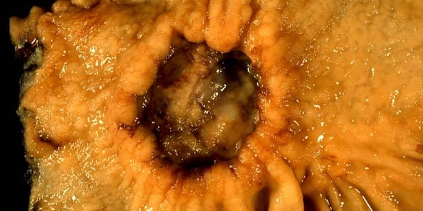
Gastric carcinoma is stomach cancer that usually begins in the stomach lining and most often forms adenocarcinoma. Symptoms may include indigestion, early satiety, weight loss, vomiting, bleeding, or anaemia; endoscopy with biopsy confirms the disease, while CT and other scans determine its stage and spread. Treatment ranges from endoscopic removal or gastric surgery to chemotherapy, immunotherapy, targeted cancer treatment, and symptom-focused care.
The best treatment plan depends on tumour location, pathology, stage, biomarkers, nutritional status, and overall fitness. Early disease may be curable, whereas advanced disease is usually managed with systemic cancer treatment and supportive care.
Key takeaways
- Adenocarcinoma is the commonest histological type.
- Helicobacter pylori is an established risk factor.
- Virchow's node (Troisier's sign), a Sister Mary Joseph nodule and Krukenberg tumours are the signs of spread to know.
- NICE NG12 refers on dysphagia at any age, or age 55 and over with weight loss and upper abdominal pain, reflux or dyspepsia.
- Staging is by CT, endoscopic ultrasound and staging laparoscopy for peritoneal disease.
What is gastric carcinoma, and how is it classified?
Gastric carcinoma is a malignant epithelial tumour that develops in the lining of the stomach. It is usually a form of stomach cancer arising from the gastric mucosa. This distinguishes it from gastric lymphoma, which develops from lymphoid tissue, and gastrointestinal stromal tumour (GIST), which arises from mesenchymal cells, often involving the muscular wall.
Gastric carcinoma is cancer originating from epithelial cells in the stomach lining. Cancer cells can invade the stomach wall, enter lymphatic vessels, and cause cancer spread to regional nodes or distant organs.
Pathology reports commonly combine histology, anatomical location, molecular findings, and TNM staging. This information helps clinicians determine whether stomach cancer can be removed surgically and which cancer treatment is most appropriate.
Histological classification
Adenocarcinoma is the commonest histological type, accounting for about 90% of stomach cancers. It is broadly divided using Lauren’s classification:
- Intestinal type: Forms glands and often produces a discrete, ulcerated or exophytic mass. It is associated with chronic Helicobacter pylori gastritis, intestinal metaplasia, and environmental risk factors.
- Diffuse type: Contains poorly cohesive cells, often including signet-ring cells. These cells contain intracellular mucin that displaces the nucleus. Extensive infiltration can produce linitis plastica, also called a “leather bottle” stomach.
Diffuse disease may be linked to inherited CDH1 mutations, which increase the risk of hereditary diffuse gastric cancer. The World Health Organization classification also recognises signet-ring cell and undifferentiated carcinomas as distinct pathological patterns.
Pathophysiology describes how abnormal cells develop, invade tissue, and produce symptoms. In intestinal-type disease, Helicobacter pylori infection may cause inflammation, atrophy, metaplasia, dysplasia, and eventually malignant transformation.
Anatomical and pathological staging
Location affects symptoms, operative planning, and prognosis. Tumours may involve the cardia, fundus, body, antrum, or pylorus. Proximal lesions may involve the gastro-oesophageal junction, while distal lesions commonly cause gastric outlet obstruction.
The gastroesophageal junction is the area where the oesophagus meets the stomach. Tumours near the gastroesophageal junction may require different surgical planning from tumours in the distal stomach, particularly when they extend into the oesophagus.
Pathological assessment records the depth of invasion, lymph-node involvement, and distant metastases. These features form the basis of TNM staging:
- T stage: Invasion through the mucosa, submucosa, muscularis, or serosa.
- N stage: Number and distribution of involved regional lymph nodes.
- M stage: Spread to organs or distant sites.
The stages describe how far the disease has progressed. Stage II gastric cancer generally indicates deeper invasion or regional lymph-node involvement without distant metastasis. Stage III gastric cancer usually reflects more extensive local invasion or nodal disease.
MRCS Part B questions may test lymphatic spread, Virchow’s node, Sister Mary Joseph nodule, or Krukenberg tumour from ovarian metastasis. Peritoneal and liver metastases also indicate advanced disease and influence treatment.
Classify stomach cancer by histology, anatomical site, depth, lymph-node spread, and distant metastasis before discussing treatment.
How does gastric carcinoma present?
Gastric carcinoma commonly causes vague upper gastrointestinal symptoms at first, while advanced disease may cause bleeding, obstruction, weight loss, or signs of cancer spread.
Early gastric carcinoma may cause few specific symptoms. The stomach can expand considerably, so cancer may remain clinically silent until it becomes advanced. Vague symptoms often resemble benign dyspepsia or peptic ulcer disease.
Symptoms of gastric cancer include indigestion, early satiety, nausea, abdominal discomfort, reduced appetite, and unexplained weight loss.
Early symptoms
Common early features include indigestion, epigastric discomfort, nausea, reduced appetite, and early satiety. Early satiety means feeling full after eating a small amount. New or persistent dyspepsia deserves investigation, and NICE NG12 sets the threshold at 55 and over with weight loss, or dysphagia at any age.
- Early stomach cancer may present with persistent dyspepsia, epigastric discomfort, nausea, reduced appetite, or early satiety.
- Early symptoms are often vague, so persistent or changing upper gastrointestinal symptoms require careful clinical assessment and appropriate investigation.
- Weight loss and abdominal pain are common presenting features when gastric cancer has progressed beyond its earliest stage.
Weight loss may result from poor intake, early satiety, cancer-related inflammation, or advanced disease. A patient may also describe fatigue or reduced exercise tolerance.
Stomach cancer diagnosed at an early stage may produce few warning signs, so symptom duration and progression matter. A person with persistent symptoms should seek medical advice rather than assume that indigestion is harmless.
Red flags and advanced presentation
The following features should prompt urgent investigation, usually with upper gastrointestinal endoscopy and biopsy. They may indicate advanced disease or a complication.
- Persistent vomiting may indicate gastric outlet obstruction, especially when accompanied by postprandial fullness, dehydration, or a visible peristaltic wave.
- Dysphagia, gastrointestinal bleeding, melaena, haematemesis, or iron-deficiency anaemia should raise suspicion of upper gastrointestinal malignancy.
- A palpable epigastric mass, marked cachexia, progressive anorexia, or rapid weight loss suggests locally advanced or metastatic stomach cancer.
The examination should assess hydration, nutritional status, abdominal tenderness, masses, organ enlargement, and lymph nodes. Succussion splash may support gastric outlet obstruction, although it is not specific.
- Virchow’s node, a hard left supraclavicular lymph node, is Troisier’s sign and suggests metastatic abdominal cancer.
- A Sister Mary Joseph nodule is a metastatic umbilical deposit, indicating possible peritoneal or intra-abdominal spread.
- Ascites and hepatomegaly may indicate peritoneal or hepatic metastases, while a palpable stomach mass suggests advanced local disease.
Symptoms can result from the primary tumour, obstruction, bleeding, inflammation, or cancer spread. Cancer spread may affect appetite, nutrition, liver function, bowel movement, and general strength.
Persistent dyspepsia becomes more concerning when it occurs with weight loss, anaemia, vomiting, bleeding, dysphagia, or progressive early satiety.
MRCS Part B clinical reasoning
For an MRCS Part B station, structure the answer around symptoms, red flags, examination, differential diagnoses, and investigations. Explain your concerns clearly, acknowledge uncertainty, and recommend urgent endoscopy with biopsy when appropriate.
Persistent dyspepsia with weight loss, anaemia, vomiting, bleeding, or metastatic signs requires urgent investigation for stomach cancer.
What Causes Stomach Cancer and Who Is at Risk?
Stomach cancer develops when abnormal cells grow uncontrollably in the stomach lining. Most stomach cancers are adenocarcinomas, arising from gland-forming cells. The cause is usually multifactorial. Infection, long-term inflammation, inherited changes, diet, and lifestyle can interact over many years.
Aetiology means the underlying causes and contributors to a disease. The aetiology of stomach cancer may include infection, chronic inflammation, inherited genetic factors, smoking, diet, age, and previous gastric surgery.
Established Causes and Risk Factors
The strongest preventable association is Helicobacter pylori infection. Persistent infection can cause chronic atrophic gastritis, which reduces normal gastric glands. This may progress to intestinal metaplasia, then gastric dysplasia, and eventually gastric carcinoma. These changes form a recognised sequence of precancerous injury.
A chronic helicobacter pylori infection can alter the stomach environment for many years. The risk from helicobacter pylori infection is higher when inflammation combines with smoking, a high-salt diet, or a family history of stomach cancer.
Other established or recognised risks include:
- A diet high in salt, smoked foods, or poorly preserved foods
- Low intake of fresh fruit and vegetables
- Tobacco use
- Heavy alcohol consumption
- Increasing age, particularly above 50 years
- Pernicious anaemia and autoimmune atrophic gastritis
- Previous stomach surgery, especially after a long interval
- A family history of stomach cancer
Previous gastric surgery can increase later risk in some patients, particularly after a long interval. The term gastric surgery includes procedures that alter the stomach’s anatomy, drainage, or exposure to bile.
People with previous gastric ulcers or chronic inflammation may also have increased risk. However, not every association proves that a factor directly causes cancer. For example, social deprivation may reflect differences in diet, infection rates, healthcare access, or smoking.
The epidemiology of disease varies by region, age, sex, diet, infection prevalence, and healthcare access. Epidemiology also helps explain why mortality can remain high where patients present with late-stage disease.
Genetic, Viral, and Other Associations
A small proportion of cases result from inherited cancer syndromes. Hereditary diffuse gastric cancer is strongly linked to pathogenic changes in the CDH1 gene, which affects cell adhesion. Carriers can develop diffuse-type disease at a younger age. Genetic counselling and specialist surveillance may be appropriate for affected families.
Inherited genetic factors may affect susceptibility, age at onset, tumour type, and treatment decisions. A strong family history should prompt discussion with a specialist genetics service.
Epstein–Barr virus is associated with a subset of gastric cancer cases. The association is recognised, but infection alone does not explain most disease. Blood group A, chronic ulceration, and some dietary preservatives have also been reported as possible contributors. Their effects are less certain than H. Pylori, smoking, or inherited CDH1 mutations.
Overall, risk reflects the combined effect of infection, inflammation, age, genetics, and exposure history. H. Pylori infection and chronic mucosal injury are major modifiable risks, while inherited CDH1 mutations identify a high-risk familial group.
Prevention still focuses on identifying and treating pylori infection, reducing tobacco exposure, and investigating persistent upper gastrointestinal symptoms. These measures may help prevent stomach cancer, although they cannot eliminate every inherited or environmental risk.
How is suspected gastric carcinoma investigated?
Endoscopy with multiple biopsies is the key test used to diagnose gastric cancer, while cross-sectional imaging determines the extent of disease and possible cancer spread.
Upper gastrointestinal endoscopy with biopsy confirms suspected gastric carcinoma. CT of the chest, abdomen, and pelvis then assesses local spread, lymph nodes, and distant metastases.
Confirming the diagnosis
Upper gastrointestinal endoscopy (OGD) is the principal diagnostic test for suspected stomach cancer. It directly visualises the mucosa and may show an ulcer, irregular mass, friable lesion, or diffuse thickening.
Multiple biopsies should be taken from the lesion and its edges. Histology confirms cancer, identifies the type, and provides tumour grading. Adenocarcinoma accounts for about 90% of gastric malignancies. Lymphoma and gastrointestinal stromal tumours are less common alternatives.
To diagnose gastric cancer, clinicians combine the patient’s history, endoscopic appearance, biopsy findings, imaging, and staging information. A biopsy identifies cancer cells and distinguishes adenocarcinoma from lymphoma, GIST, or another tumour.
A normal-looking area does not always exclude disease. Diffuse infiltrative cancer, such as linitis plastica, can produce a stiff, poorly distensible “leather bottle” stomach. Superficial biopsies may miss deeper disease, so repeat sampling or further assessment may be required.
Endoscopy also helps assess bleeding, gastric outlet obstruction, and the tumour’s distance from the gastro-oesophageal junction. These findings influence the surgical approach and whether resection might be curative.
Staging investigations
Contrast-enhanced CT of the chest, abdomen, and pelvis is the standard staging scan. It assesses the primary tumour, regional lymph nodes, liver, lungs, peritoneum, and other sites of metastatic disease.
Staging determines whether the stomach cancer is localised, regional, or metastatic. Stage IV gastric cancer means that distant metastatic disease is present, while stage IV gastric disease generally requires systemic treatment rather than surgery alone.
Surgeons should look for gastric wall thickening, perigastric fat invasion, enlarged nodes, ascites, liver lesions, and peritoneal nodules. CT may also identify an ovarian mass from transcoelomic spread, known as a Krukenberg tumour. Mucinous lesions can have low attenuation and may contain calcification.
Endoscopic ultrasound (EUS) provides high-resolution assessment of the stomach wall. It helps estimate the depth of invasion and may assess nearby lymph nodes. EUS-guided needle sampling can confirm suspicious nodes when the result would alter treatment.
Diagnostic or staging laparoscopy is useful when peritoneal or serosal spread remains possible despite CT. The surgeon inspects the abdominal cavity and may obtain peritoneal biopsies or washings for cytology. Positive peritoneal cytology indicates metastatic disease and may prevent curative gastrectomy.
PET-CT can identify metabolically active distant disease in selected patients. Its sensitivity is limited in some mucinous and diffuse cancers, so a negative scan does not exclude metastases.
When cancer spreads, imaging may show liver lesions, lung nodules, peritoneal deposits, enlarged nodes, or bone disease. Cancer spreads through direct invasion, lymphatic channels, blood vessels, or transcoelomic movement across the peritoneum.
Blood tests and tumour markers
Symptoms of initial blood tests include a full blood count, liver function tests, renal function, electrolytes, and clotting studies. The full blood count may show iron-deficiency anaemia from chronic blood loss. Liver tests can suggest hepatic metastases or provide a baseline before treatment.
Symptoms of nutritional assessment should include weight loss, body mass index, oral intake, and functional status. Albumin may support nutritional and prognostic assessment, although inflammation also lowers it. Cancer-related cachexia can affect surgical fitness and recovery.
Tumour markers, such as CEA and CA 19-9, are not reliable diagnostic tests. They may be measured selectively as baseline values and during follow-up. No single blood test can confirm or exclude stomach cancer.
Endoscopy with adequate biopsy establishes the diagnosis, while CT and selective laparoscopy determine whether treatment can be curative.
How is gastric carcinoma treated?
Treatment for stomach cancer depends on stage, resectability, tumour biology, symptoms, and the patient’s goals and fitness.
What determines treatment?
Treatment depends on tumour stage, location, lymph-node involvement, distant spread, resectability, and the patient’s fitness. CT, diagnostic laparoscopy, biopsy results, and biomarker testing help define the plan.
A specialist multidisciplinary team (MDT) should review each case. The team usually includes upper gastrointestinal surgeons, oncologists, radiologists, pathologists, specialist nurses, and dietitians.
A curative operation requires complete removal of the tumour with clear margins and appropriate lymph-node clearance. Frailty, nutrition, organ function, and patient preferences also influence the decision.
A treatment option overview should include endoscopic therapy, surgery, perioperative chemotherapy, immunotherapy, targeted drugs, radiotherapy, and palliation. The most suitable treatment option changes when cancer cells have caused distant cancer spread.
What surgery is used?
Endoscopic resection may treat carefully selected early lesions. It is suitable when the tumour is superficial, small, and has a low risk of lymph-node spread. Endoscopic submucosal dissection can remove some T1a lesions without removing the stomach.
For invasive but localised disease, surgeons may perform a subtotal or total gastrectomy:
- Subtotal gastrectomy: removes part of the stomach, usually for distal tumours.
- Total gastrectomy: removes the whole stomach, often for proximal, extensive, or diffuse disease.
- Lymphadenectomy: removes regional nodes for staging and cancer control. A D2 dissection is commonly performed in specialist centres.
- Reconstruction: restores gastrointestinal continuity. After total gastrectomy, a Roux-en-Y oesophagojejunostomy connects the oesophagus to the small bowel.
The operation selected for gastric carcinoma must balance oncological clearance with preservation of function. A total gastrectomy is not automatically better than a subtotal procedure if both achieve clear margins.
Surgery may be a standard treatment for localised disease, but it is rarely the sole standard treatment for stage iv gastric cancer. If cancer spread is extensive, systemic cancer treatment may provide greater benefit than a major operation.
How does systemic therapy help?
Perioperative chemotherapy is often used for resectable, locally advanced stomach cancer. It may shrink the tumour before surgery and treat microscopic metastatic disease afterward. A commonly used approach is a platinum-fluoropyrimidine regimen, such as FLOT, when the patient can tolerate intensive therapy.
Chemoradiotherapy has a more selective role. It may be considered after an incomplete resection, for positive margins, or in specific treatment pathways. It can also help control local symptoms when surgery is unsuitable.
Advanced tumours should undergo biomarker testing. HER2-positive disease may benefit from trastuzumab combined with systemic chemotherapy. PD-L1 expression, mismatch-repair status, and microsatellite instability can guide immunotherapy, including immune checkpoint inhibitors. Tumours with MSI-high or deficient mismatch repair may respond particularly well to immunotherapy. In specific advanced disease settings, agents such as Tevimbra (tislelizumab) may also be considered. Other actionable changes, including NTRK, BRAF, or RET alterations, may support targeted treatment.
The standard treatment for IV gastric cancer is usually biomarker-guided systemic therapy, although selected patients may receive local procedures. In stage IV gastric cancer, cancer cells may have reached the liver, lungs, peritoneum, distant lymph nodes, or another organ.
What happens when the cancer cannot be removed?
Unresectable or metastatic disease is usually managed with palliative systemic treatment. Options include chemotherapy, targeted drugs, immunotherapy, or later-line agents such as trifluridine and tipiracil.
When stomach cancer spreads, the aim of cancer treatment may shift from cure to disease control, symptom relief, and quality of life. Patients with stage IV gastric disease should still receive an individualised treatment option based on biomarkers and fitness.
Treatment should also relieve specific problems. An endoluminal stent can bypass malignant gastric outlet obstruction. Feeding jejunostomy or another enteral feeding route may support nutrition. A gastrojejunostomy can provide a surgical bypass for selected, fitter patients.
Radiotherapy may reduce bleeding, pain, or obstruction. Palliative resection is reserved for carefully selected patients with persistent bleeding or obstruction - NCI).
What complications follow treatment?
Symptoms of early surgical complications include anastomotic leak, bleeding, infection, ileus, and pulmonary complications. Later nutritional effects include weight loss, early satiety, diarrhoea, and iron or folate deficiency.
Dumping syndrome causes abdominal cramps, sweating, dizziness, or diarrhoea after meals. Smaller meals and limiting rapidly absorbed sugars can help. Bile reflux may cause epigastric discomfort, nausea, and vomiting.
After total gastrectomy, patients cannot absorb vitamin B12 normally because the stomach no longer produces intrinsic factor. Lifelong vitamin B12 replacement is usually required, with regular monitoring of weight, iron, folate, calcium, and vitamin D.
Gastric carcinoma treatment is stage-led and MDT-based: remove resectable disease when feasible, use biomarker-guided systemic therapy, and prioritise symptom control when cure is not possible.
Treatment for gastric carcinoma is tailored to tumour stage, resectability, spread, biomarkers, overall fitness, and patient preferences. This quick-reference table summarises the main approaches, from potentially curative local treatment to systemic therapy and palliation within a specialist multidisciplinary team.
In the 2026 treatment landscape, a standard treatment may be modified by HER2, PD-L1, MSI, mismatch-repair, or other molecular results. Another standard treatment may be unsuitable if nutrition, organ function, or performance status is poor.
A treatment option for localised disease may be curative, while a treatment option for metastatic disease often aims to control cancer spread. The phrase stomach cancer treated therefore describes several different pathways rather than one universal operation.
What should you know about prognosis for the exam?
Gastric carcinoma is a malignant tumour arising from the stomach, most often adenocarcinoma. Prognosis depends on stage, tumour biology, response to treatment, nutritional status, and whether cancer spread has occurred. Factors affect prognosis by influencing resectability, lymph-node burden, distant metastases, and response to systemic therapy.
Prognosis and prevention
Prognosis depends mainly on stage at diagnosis. Tumour depth, lymph-node involvement, and distant metastases strongly influence survival. Early stomach cancer has a better outlook because it is less likely to involve lymph nodes and may be cured by resection.
The mortality of stomach cancer is strongly linked to late presentation and unequal access to endoscopy, staging, surgery, and systemic therapy. Mortality statistics vary by country and by stage distribution.
Histological subtype also matters. Diffuse-type disease, including linitis plastica, can spread widely through the stomach wall. Response to chemotherapy, targeted treatment, immunotherapy, and surgery further affects outcome. Treatment plans should follow current specialist guidance, such as the NCCN recommendations.
Prevention focuses on reducing avoidable risks. Test for and eradicate Helicobacter pylori when indicated. Stop smoking, limit alcohol, and maintain a balanced diet with fewer smoked, salted, or preserved foods. Chronic atrophic gastritis and hereditary syndromes may require specialist assessment.
Eradicating helicobacter pylori infection may help prevent stomach cancer, especially before irreversible intestinal metaplasia develops. Testing for a persistent pylori infection may involve breath, stool, blood, or biopsy-based methods.
There is no universal population screening programme for stomach cancer in the UK. However, high-risk patients may need genetic counselling, endoscopic surveillance, or risk-reducing surgery. New, persistent dyspepsia, weight loss, anaemia, vomiting, or gastrointestinal bleeding requires clinical assessment.
A balanced diet, smoking cessation, appropriate infection treatment, and specialist surveillance can help prevent stomach cancer. These measures do not guarantee prevention because some cancer cells arise through inherited or incompletely understood mechanisms.
MRCS Part B revision priorities
Common applied questions include:
- Virchow’s node: a left supraclavicular node suggesting advanced metastatic spread.
- Sister Mary Joseph nodule: an umbilical deposit from intra-abdominal cancer.
- Krukenberg tumour: ovarian metastasis, classically from diffuse gastric cancer.
- Gastric lymphatic drainage and the importance of regional lymphadenectomy.
- The anatomy of subtotal and total gastrectomy, including the oesophagus, duodenum, pancreas, spleen, and major vessels.
- Surgical complications, such as anastomotic leak, bleeding, infection, dumping syndrome, bile reflux, and nutritional deficiencies.
For revision, remember that cancer spreads by direct invasion, lymphatics, blood vessels, and transcoelomic routes. Cancer spread may produce Virchow’s node, a Sister Mary Joseph nodule, Krukenberg tumour, liver metastasis, or peritoneal deposits.
The MRCS Part B includes two surgical pathology stations. Each station is marked out of 20, with all marks awarded for clinical knowledge and its application. Practise explaining pathology, staging, investigations, treatment, and complications aloud.
Mrcspartbquestions provides more than 3,500 interactive questions, pathology explanations, anatomy spot tests, examination scenarios, and surgical skills practice. Use mark schemes, notes, and progress tracking to identify weak areas across devices.
What is the highest-yield sign of advanced stomach cancer? Virchow’s node, although it is not present in every patient. Early detection, accurate staging, and focused practice of anatomy, metastatic signs, and surgical complications are central to gastric carcinoma outcomes and MRCS Part B success.
How is this topic marked in MRCS Part B?
MRCS Part B marks a surgical pathology station out of 20, with all 20 marks for clinical knowledge and its application. Two of the seventeen examined stations are surgical pathology, the second titled surgical pathology and/or microbiology.