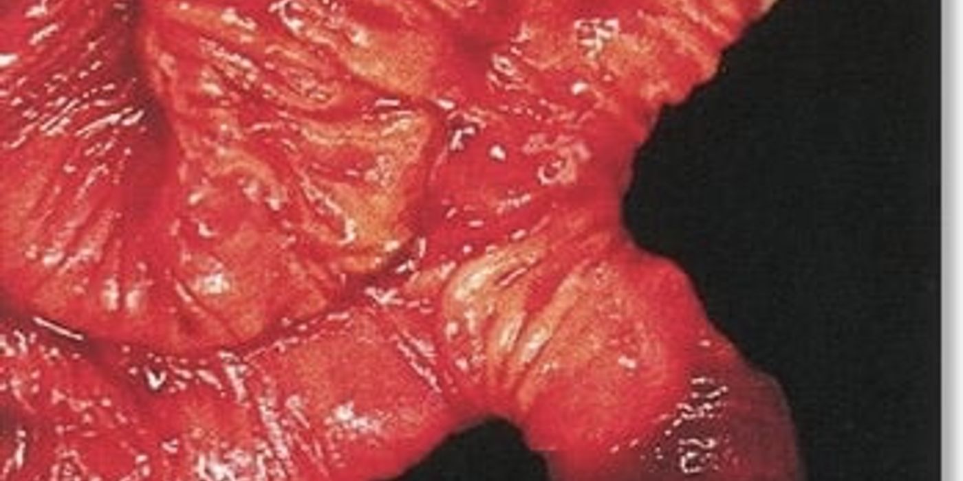
Meckel’s diverticulum is understood as a congenital true diverticulum of the ileum caused by persistence of the omphalomesenteric duct. It is usually symptom-free, but a complicated Meckel diverticulum can cause gastrointestinal haemorrhage, inflammation, intussusception, perforation, or obstruction and may require urgent surgery.
This 2026 guide explains how Meckel diverticulum occurs, why ectopic stomach tissue causes ulceration, how clinicians diagnose it, and when diverticulectomy or ileal resection is needed.
Key takeaways
- The rule of 2s: about 2% of the population, 2 feet from the ileocaecal valve, 2 inches long, 2 types of ectopic tissue, and commonly presenting before the age of 2.
- It is a true diverticulum, containing all layers of the bowel wall, and a remnant of the vitellointestinal duct.
- A technetium-99m pertechnetate scan detects ectopic gastric mucosa.
- It presents with bleeding, obstruction, diverticulitis or intussusception.
- It is the commonest congenital anomaly of the gastrointestinal tract.
- StatPearls puts ectopic gastric mucosa in about 60% of diverticula; a pertechnetate scan enhanced with cimetidine has a reported sensitivity of 85% and specificity of 95%.
What is a Meckel's diverticulum?
Meckel’s diverticulum is a congenital outpouching of the small intestine that contains the full thickness of the intestinal wall. Meckel's diverticulum is a true congenital diverticulum arising from the antimesenteric border of the ileum.
A true diverticulum contains all layers of the bowel wall. This distinguishes it from a false diverticulum, which lacks one or more wall layers. The pouch usually lies within about 2 feet of the ileocaecal valve.
Meckel diverticulum is a persistent embryological remnant that projects from the ileum, the final part of the small intestine. Unlike acquired diverticula, it forms before birth and is not primarily caused by raised intestinal pressure.
Embryology and anatomy
This condition results from incomplete obliteration of the omphalomesenteric duct, also called the vitellointestinal duct. During early development, this duct connects the embryonic midgut to the yolk sac. Persistence of part of the duct leaves an ileal outpouching.
The yolk stalk normally disappears as the umbilical and intestinal structures mature. Persistence of the yolk stalk can leave a fibrous cord, an umbilical sinus, or a connection between the intestine and the umbilicus.
The diverticulum may contain ectopic, or misplaced, tissue. Gastric mucosa is the most common type. It can secrete acid, causing adjacent ileal ulceration and gastrointestinal bleeding. Pancreatic tissue may also occur.
Heterotopic tissue is normal tissue located in an abnormal anatomical site. In Meckel’s diverticulum, heterotopic stomach tissue or pancreatic tissue can alter symptoms, scan results, and operative risk.
The remnant can have a narrow or broad stalk. Its blood supply may arise from an ileal branch of the superior mesenteric artery, and an associated fibrous cord can tether the intestine to the umbilicus or mesentery.
The rule of twos
The classic teaching aid is the rule of twos:
- Affects about 2% of the population
- Measures approximately 2 inches long
- Lies within 2 feet of the ileocaecal valve
- Contains two common types of ectopic tissue: gastric and pancreatic
- Commonly presents before 2 years of age
- Symptomatic cases are more common in males, traditionally described as a 2:1 ratio
These figures support recall during an MRCS Part B station. However, they are approximations, not diagnostic rules. Many patients remain asymptomatic throughout life. Location, size, tissue type, and age can vary considerably.
The incidence is commonly estimated at approximately 2%, although reported incidence varies by study population and method of detection. An incidentally discovered lesion may never have caused symptoms, while a complicated Meckel diverticulum is more likely to be found during emergency imaging or operation.
Clinical significance
Complications are more common in children because symptomatic disease often appears early. Bleeding, inflammation, obstruction, intussusception, and volvulus may occur. Inflammation can closely mimic acute appendicitis. However, complications can present at any age, including adulthood.
Symptomatic patients may require surgery to remove the diverticulum or affected ileum. Asymptomatic incidental findings usually do not require treatment, although management depends on individual risk factors.
The incidence of symptoms is influenced by age, sex, diverticulum anatomy, and the presence of heterotopic stomach tissue. In paediatric patients, the risk of a bleeding Meckel diverticulum is particularly important because acid-producing tissue may ulcerate the adjacent ileum.
Clinical insight: A normal-looking umbilicus does not exclude a persistent yolk stalk remnant. The relevant lesion is usually attached to the antimesenteric ileum, not directly to the umbilicus.
Why is this condition tested in the MRCS Part B? Its embryology, anatomy, complications, and surgical treatment link directly to applied surgical pathology and clinical decision-making.
Meckel's diverticulum is a persistent omphalomesenteric duct remnant and a true ileal diverticulum that can cause bleeding, obstruction, or inflammation at any age.
What Are the Symptoms of Meckel's Diverticulum?
Meckel’s diverticulum is often asymptomatic, but symptoms develop when ectopic tissue ulcerates the ileum or when the pouch becomes inflamed, trapped, or twisted.
Most people with a Meckel diverticulum have no symptoms. Only about 2% of people develop symptomatic disease, although complications can be serious. Symptoms depend on bleeding, inflammation, or impaired bowel movement.
- Painless, dark-red rectal bleeding is the classic presentation when ectopic gastric tissue causes ileal ulceration.
- Children commonly present with sudden, substantial bleeding, while adults more often develop abdominal pain, obstruction, or inflammation.
- Abdominal pain with guarding, fever, vomiting, or distension suggests diverticulitis, perforation, or another acute complication.
- Intussusception occurs when the diverticulum acts as a lead point, causing colicky pain, vomiting, and sometimes bloody stool.
- Volvulus or an adhesive band can twist or compress the small bowel, producing obstruction and progressive abdominal distension.
- Perforation causes severe, localised or generalised pain, abdominal rigidity, tachycardia, and clinical signs of peritonitis.
- Ongoing or repeated blood loss may cause iron-deficiency anaemia, fatigue, pallor, dizziness, or shortness of breath.
- A Meckel diverticulum complication requires urgent assessment when bleeding, severe pain, persistent vomiting, or shock develops.
In meckel diverticulum children, the most useful clinical clues are painless maroon stool, unexplained anaemia, episodic abdominal symptoms, or an acute abdomen. A paediatric or paediatric surgical team should assess a child with substantial rectal blood loss.
The incidence of symptomatic disease is highest in paediatric and young-adult groups, although an incidentally discovered remnant can remain silent for decades. A doctor should consider the diagnosis when common explanations do not fit the pattern.
Bleeding symptoms
The most characteristic symptom is painless lower gastrointestinal bleeding. Ectopic gastric mucosa secretes acid, which can ulcerate the adjacent ileal lining. The diverticulum itself may not become inflamed, so bleeding can occur without pain.
Children may pass a large volume of dark-red blood. Brick-red, jelly-like stool can also occur. Adults may develop melaena, which appears as black, tarry stool. Continued haemorrhage can cause anaemia and circulatory instability.
A bleeding Meckel diverticulum can be intermittent, making diagnosis difficult between episodes. The blood may originate from an ulcer in the adjacent small intestine rather than from the diverticulum’s lining itself.
The quantity of blood does not reliably indicate the size of the pouch. A small ulcer near an artery can produce substantial haemorrhage, while a larger lesion may remain clinically silent.
Pain and obstructive complications
Painful presentations usually reflect inflammation or mechanical obstruction. Diverticulitis may resemble appendicitis, causing right lower-quadrant pain, tenderness, fever, nausea, and vomiting. Perforation can progress to peritonitis and sepsis.
Obstruction may result from intussusception, volvulus, an inflammatory mass, or a fibrous vitelline duct band. Patients can develop cramping pain, vomiting, constipation, and increasing distension. These complications may require emergency surgery.
In children, bleeding is the commonest presentation, often before age two. A child with severe abdominal pain, vomiting, distension, or bloody stool may have intussusception or obstruction. In adults and older patients, diverticulitis, perforation, anaemia, and bowel obstruction are more frequent clinical concerns.
A fibrous cord can connect the diverticulum to the umbilicus or mesentery. Small intestine may pass beneath this cord, creating a closed loop. The stalk can also act as a lead point for telescoping of the intestine.
The symptoms can overlap with appendicitis, peptic ulceration, inflammatory bowel disease, and other causes of gastrointestinal bleeding. Clinical assessment must therefore identify complications early and guide treatment, imaging, endoscopy, or surgery.
A Meckel diverticulum most often causes painless bleeding in children, but pain, obstruction, inflammation, and perforation can occur at any age.
What is the embryology, and why does ectopic tissue matter?
Meckel’s diverticulum results from persistence of the embryonic yolk stalk, also called the vitelline or omphalomesenteric duct. The underlying problem is incomplete embryological development. Meckel’s diverticulum is a true diverticulum caused by failure of the vitelline, or omphalomesenteric, duct to disappear. This duct connects the developing midgut to the yolk sac. It normally obliterates during the fifth week of fetal development. If it remains partly open, an ileal pouch forms on the antimesenteric border.
The causes Meckel condition is therefore developmental rather than infectious or dietary. Meckel diverticulum occurs when persistence of the yolk stalk or related duct leaves tissue attached to the intestine.
How the diverticulum forms
The direct answer is that persistent duct tissue creates a congenital outpouching of the distal small bowel. Unlike a false diverticulum, this pouch contains all layers of the intestinal wall. Meckel diverticulum is therefore a remnant of fetal development, rather than an acquired lesion caused by pressure or inflammation.
The pouch may contain ectopic tissue, meaning normal tissue located in an abnormal site. Gastric mucosa is the most clinically significant type. Pancreatic tissue occurs less often.
The yolk sac supplies early embryonic nutrition. The yolk stalk connects that sac with the developing intestine before the umbilical circulation and placenta become fully established. Persistence of the stalk can produce several anatomical anomalies, including a fibrous cord or an umbilical connection.
The intestinal wall remains continuous with the diverticulum. This explains why a true diverticulum can contain mucosa, submucosa, muscularis, and serosa, and why inflammation may extend into surrounding ileum.
Why ectopic tissue causes complications
Ectopic gastric mucosa secretes hydrochloric acid. The adjacent ileum does not have the same protective mechanisms as the stomach. Acid exposure can therefore produce peptic ulceration in nearby ileal mucosa. This ulcer may erode blood vessels and cause painless or severe lower gastrointestinal bleeding.
Stomach tissue within the pouch may be clinically silent, but acid-producing stomach tissue beside unprotected ileal mucosa can create ulceration. Heterotopic pancreatic tissue may also be found, although it is less often responsible for the presenting symptoms.
Ulceration can also cause inflammation, perforation, and local peritonitis. The diverticulum may become inflamed, producing diverticulitis that resembles appendicitis. A fibrous band can cause volvulus or small bowel obstruction. The pouch can also act as a lead point for intussusception. These complications explain why treatment differs between incidental and symptomatic disease.
Risk is higher in males, younger patients, and people with ectopic tissue. Symptoms commonly occur during childhood, although adults can present with bleeding, obstruction, or inflammation. The classic rule of twos remains useful for examinations: 2% of people are affected, the pouch is about 2 inches long, and it lies about 2 feet from the ileocaecal valve. Symptomatic presentation is traditionally associated with age two, but this is only an approximate teaching rule.
A persistent fibrous cord may extend from the diverticulum toward the umbilicus. This cord can compress the small intestine, create a volvulus, or form an internal hernia. An artery accompanying the cord may also increase operative complexity.
Persistent vitelline duct tissue creates the pouch, while ectopic gastric mucosa drives ulceration, bleeding, and major complications.
How Is Meckel's Diverticulum Diagnosed?
Diagnosis depends on the clinical presentation, age, stability, and suspected complication; no single test detects every lesion. Diagnosis begins with clinical assessment, abdominal examination, and blood tests. In children with unexplained gastrointestinal bleeding, a technetium-99m pertechnetate Meckel scan is usually the key investigation, while CT, endoscopy, angiography, or surgery may be needed in selected cases.
Initial assessment
Diagnosis starts with the patient’s age, symptoms, and clinical stability. Ask about rectal bleeding, melaena, abdominal pain, vomiting, fever, bowel obstruction, and previous episodes. Severe bleeding, guarding, shock, or peritonitis requires urgent resuscitation and surgical review.
Abdominal examination may show tenderness, guarding, distension, or a palpable mass. Localised right lower-quadrant pain can resemble appendicitis. Colicky pain and vomiting raise concern for obstruction or intussusception caused by an inverted diverticulum.
Blood tests help assess the effect and possible cause of the presentation. Full blood count may show anaemia from chronic or acute haemorrhage. Haemoglobin can initially appear normal after sudden blood loss, so repeat measurement may be needed. White cell count and C-reactive protein may support inflammation, diverticulitis, perforation, or another intra-abdominal process.
Urea, electrolytes, creatinine, liver tests, coagulation studies, and group-and-save testing help prepare for contrast imaging, endoscopy, or surgery. A falling haemoglobin with painless rectal bleeding supports a bleeding source, but does not confirm a Meckel diverticulum.
The incidence of a positive scan depends on the amount of functioning stomach tissue and the timing of the study. A doctor may therefore combine laboratory results with ultrasound, CT, endoscopy, or operative findings.
Meckel scan and imaging
A Meckel scan uses technetium-99m pertechnetate, which is taken up by ectopic gastric mucosa. This makes the scan particularly useful for children with unexplained gastrointestinal bleeding. It is non-invasive and can identify functioning gastric tissue within the pouch.
The scan has limitations. It may miss a small lesion, low-volume bleeding, or a diverticulum without sufficient gastric mucosa. Results can also be affected by bowel activity and previous bleeding. A negative scan does not exclude the condition, especially in adults.
CT abdomen and pelvis is useful when pain, obstruction, inflammation, perforation, or an alternative diagnosis is suspected. It may show a blind-ending, fluid-filled structure arising from the ileum. However, a normal or unclear CT does not reliably exclude a small diverticulum.
Ultrasound is radiation-free and useful in children. It may identify intussusception, an obstructed bowel loop, or an inflamed tubular structure. Its accuracy depends on operator experience, bowel gas, and the patient’s body habitus.
An incidentally discovered Meckel diverticulum may be visible during CT, laparoscopy, or unrelated abdominal surgery. The incidence of incidental identification increases when modern imaging and minimally invasive procedures provide broader views of the abdomen.
Selected investigations and differential diagnosis
Endoscopy can exclude upper gastrointestinal bleeding, peptic ulcer disease, and some colonic causes of haemorrhage. Capsule endoscopy or double-balloon enteroscopy may assess the small bowel when routine tests are unrevealing, although capsule retention is a concern during obstruction.
Mesenteric angiography can locate active bleeding and may allow embolisation. It is most useful during significant ongoing haemorrhage, but it may be negative when bleeding is intermittent. Diagnostic laparoscopy provides direct inspection and can proceed to resection when symptoms strongly suggest a surgical cause.
The differential diagnosis depends on the presentation. Appendicitis usually causes progressive right iliac fossa pain and inflammatory markers. Crohn disease may cause diarrhoea, weight loss, anaemia, and terminal ileal inflammation. Intussusception causes episodic colicky pain and obstruction. Peptic ulcer disease commonly causes melaena or upper abdominal symptoms. Other causes include inflammatory bowel disease, polyps, vascular malformations, and gastrointestinal infection.
A practical diagnostic pathway combines clinical findings, haemoglobin and inflammatory markers, targeted Meckel scanning, cross-sectional imaging, and surgery when complications or persistent bleeding demand it.
Clinicians may use electronic health records, radiology workstations, and structured paediatric haemorrhage pathways to reduce diagnostic delay. These tools support, but do not replace, examination and senior surgical judgement.
How is a Meckel's diverticulum treated?
Treatment depends on whether the lesion is symptomatic, complicated, or found incidentally during another procedure.
What is the initial treatment?
Emergency treatment begins with resuscitation, source control, and early surgical review. Patients with major bleeding, obstruction, perforation, or sepsis need an acute surgical approach.
- Use an ABCDE assessment and give oxygen if required.
- Insert two large-bore intravenous cannulas.
- Send full blood count, electrolytes, lactate, coagulation studies, and group-and-crossmatch samples.
- Give intravenous fluids and blood products for haemorrhagic shock.
- Insert a urinary catheter to monitor output.
- Use a nasogastric tube for persistent vomiting or bowel obstruction.
- Give broad-spectrum intravenous antibiotics when perforation, ischaemia, or sepsis is suspected.
Urgent contrast CT can help define obstruction, perforation, or an inflamed diverticulum in a stable patient. However, shock, peritonitis, uncontrolled bleeding, or strangulated obstruction requires prompt operation. Resuscitation must continue during transfer to theatre.
The treatment Meckel diverticulum pathway should be led by a general surgeon, paediatric surgeon, anaesthetist, and radiologist when appropriate. The exact plan depends on haemodynamic status, age, blood loss, imaging, and the condition of the surrounding ileum.
Why is surgery required?
Symptomatic disease generally requires removal. Indications include recurrent or severe bleeding, diverticulitis, perforation, intussusception, volvulus, obstruction from a mesodiverticular band, and suspected malignancy.
For bleeding, the surgeon must inspect the adjacent ileum. An ulcer in nearby ileum may contain ectopic gastric mucosa and can continue bleeding after simple removal of the pouch. The ulcerated segment should therefore be included in a small-bowel resection.
How is the operation selected?
A diverticulectomy removes the pouch at its base. It is suitable for a long, narrow diverticulum with simple inflammation, active bleeding without adjacent ileal ulceration, or a suitable incidental finding.
A wedge resection removes the diverticulum and a small cuff of ileum. Segmental small-bowel resection removes the diverticulum with adjoining ileum and requires an anastomosis. This approach is preferred when the base is broad, the bowel is inflamed or perforated, the diverticulum is short, or adjacent ulceration, ischaemia, or tumour is present.
An ileal resection may be safer than isolated diverticulectomy when the intestinal wall is ulcerated, inflamed, perforated, or supplied by an abnormal artery. A second ileal resection may be required if the first anastomosis would leave compromised tissue.
The surgeon should run the small bowel and identify the mesodiverticular band. This band may contain a persistent vitelline artery and cause volvulus or obstruction. It must be divided safely, with careful control of bleeding.
What are the operative approaches?
Laparoscopic surgery offers smaller wounds, less postoperative pain, and faster mobilisation. The surgeon can inspect the abdomen, identify the diverticulum, assess the mesodiverticular band, and exteriorise bowel through a small incision if needed.
Open surgery remains appropriate for haemodynamic instability, gross contamination, severe distension, extensive adhesions, or uncertain anatomy. Both approaches allow stapled or hand-sewn closure. Stapling can shorten operative time and avoids opening the bowel lumen.
An incidentally discovered lesion does not always require removal. The decision depends on age, operative risk, diverticulum length and width, inflammation, palpable ectopic tissue, a fibrous band, and the appearance of the bowel. Removal is more reasonable in a young patient with low operative risk. It may be safer to leave an uncomplicated lesion in an older or high-risk patient.
Minimally invasive techniques such as laparoscopic and robotic platforms may support inspection and resection in selected patients. Availability, surgeon experience, equipment costs, and the patient’s stability influence platform selection.
What happens after surgery?
Postoperative care includes analgesia, fluid and electrolyte replacement, venous thromboembolism prevention, early mobilisation, and observation for ileus, anastomotic leak, wound infection, intra-abdominal abscess, bleeding, and recurrent obstruction. Histology should confirm all bowel-wall layers and identify ectopic gastric or pancreatic tissue, ulceration, inflammation, or malignancy.
Uncomplicated laparoscopic recovery often allows discharge within 2–5 days. Open surgery or bowel resection may require a longer admission. The key MRCS answer is: stabilise first, remove the diverticulum, and resect adjacent ileum when the base or ulcerated bowel is diseased.
Symptoms of potential side effects include wound infection, ileus, anastomotic leakage, adhesions, recurrent obstruction, and adverse reactions to anaesthesia or medication. A doctor should explain these risks before elective intervention.
This quick-reference table summarises initial meckel's diverticulum treatment, highlighting the emergency priorities of resuscitation, diagnostic assessment, infection control, monitoring, and early surgical review for patients with bleeding, obstruction, perforation, or sepsis.
What complications does it cause, and what will you be asked?
The complications Meckel diverticulum can produce include haemorrhage, diverticulitis, perforation, intussusception, volvulus, and intestinal obstruction. Meckel’s diverticulum is a true ileal diverticulum caused by incomplete obliteration of the vitellointestinal duct. It contains all bowel-wall layers and usually arises from the antimesenteric border. Most cases remain silent, but complications may require urgent surgery.
Complications to prioritise
The main complications are bleeding, diverticulitis, perforation, small-bowel obstruction, intussusception, and volvulus. Rarely, neoplasia develops within the diverticulum. Only about 2% of people with this congenital remnant become symptomatic, although complication rates vary between studies.
Ectopic gastric mucosa can secrete acid, causing adjacent ileal ulceration and significant painless bleeding. A technetium-99m pertechnetate scan may identify this tissue, particularly in children. The classic teaching is increased tracer uptake in ectopic gastric mucosa.
Inflammation causes right iliac fossa pain, fever, vomiting, and guarding. Perforation may resemble appendicitis or cause generalised peritonitis. A diverticulum can form a lead point for intussusception. Adhesions, a mesodiverticular band, volvulus, or herniation can cause obstruction.
Management depends on the presentation. Resuscitate bleeding patients, assess haemodynamic status, and involve senior surgical and anaesthetic teams. Suspected perforation, strangulation, or persistent obstruction requires urgent surgery. Treatment may involve diverticulectomy, segmental ileal resection, anastomosis, or removal of a band. Incidental resection remains a risk–benefit decision.
A complicated Meckel diverticulum can compromise the blood supply of adjacent ileum. The artery supplying the stalk or cord should be identified before division, particularly when inflammation distorts normal anatomy.
MRCS Part B clinical scenarios
Practise answering these station prompts aloud:
- A child has painless melaena: ask about bleeding, pain, vomiting, and previous episodes. Assess airway, breathing, circulation, perfusion, and haemoglobin. Consider ectopic gastric mucosa and arrange specialist imaging.
- An adult has right iliac fossa pain: examine for tenderness, guarding, masses, and peritonism. Your differential includes appendicitis, diverticulitis, Crohn’s disease, and perforated ulcer.
- A patient has colicky pain and vomiting: ask about distension, stool, flatus, and previous surgery. Examine for dehydration, scars, bowel sounds, hernias, and peritonitis. Start obstruction management and seek urgent surgical review.
Paediatric candidates should remember that meckel diverticulum children may present with an acute abdomen, but painless rectal blood is a particularly important clue. In paediatric emergency practice, resuscitation takes priority over completing every diagnostic test.
Applied anatomy and revision strategy
Know the rule of twos: 2% prevalence, 2 inches long, 2 feet from the ileocaecal valve, two common ectopic tissues, and presentation before age two. The vitelline artery, usually related to the superior mesenteric circulation, may persist and create operative bleeding risk. Look for an antimesenteric pouch, inflamed tip, ulceration, perforation, or a fibrous band during surgery.
Mrcspartbquestions provides interactive questions, explanations, anatomy spot tests, and surgical scenarios. Use progress tracking across desktop, tablet, and mobile devices to target weak areas.
A useful revision comparison is “bleeding in a young child, inflammation in an adult, and obstruction at any age.” This does not replace clinical reasoning, but it helps connect embryology, anatomy, symptoms, imaging, and operative management.
MRCS candidates should link the diverticulum’s embryology and anatomy to bleeding, obstruction, inflammation, and the need for timely surgery.
How is this topic marked in MRCS Part B?
MRCS Part B marks a surgical pathology station out of 20, with all 20 marks for clinical knowledge and its application. Two of the seventeen examined stations are surgical pathology, the second titled surgical pathology and/or microbiology.