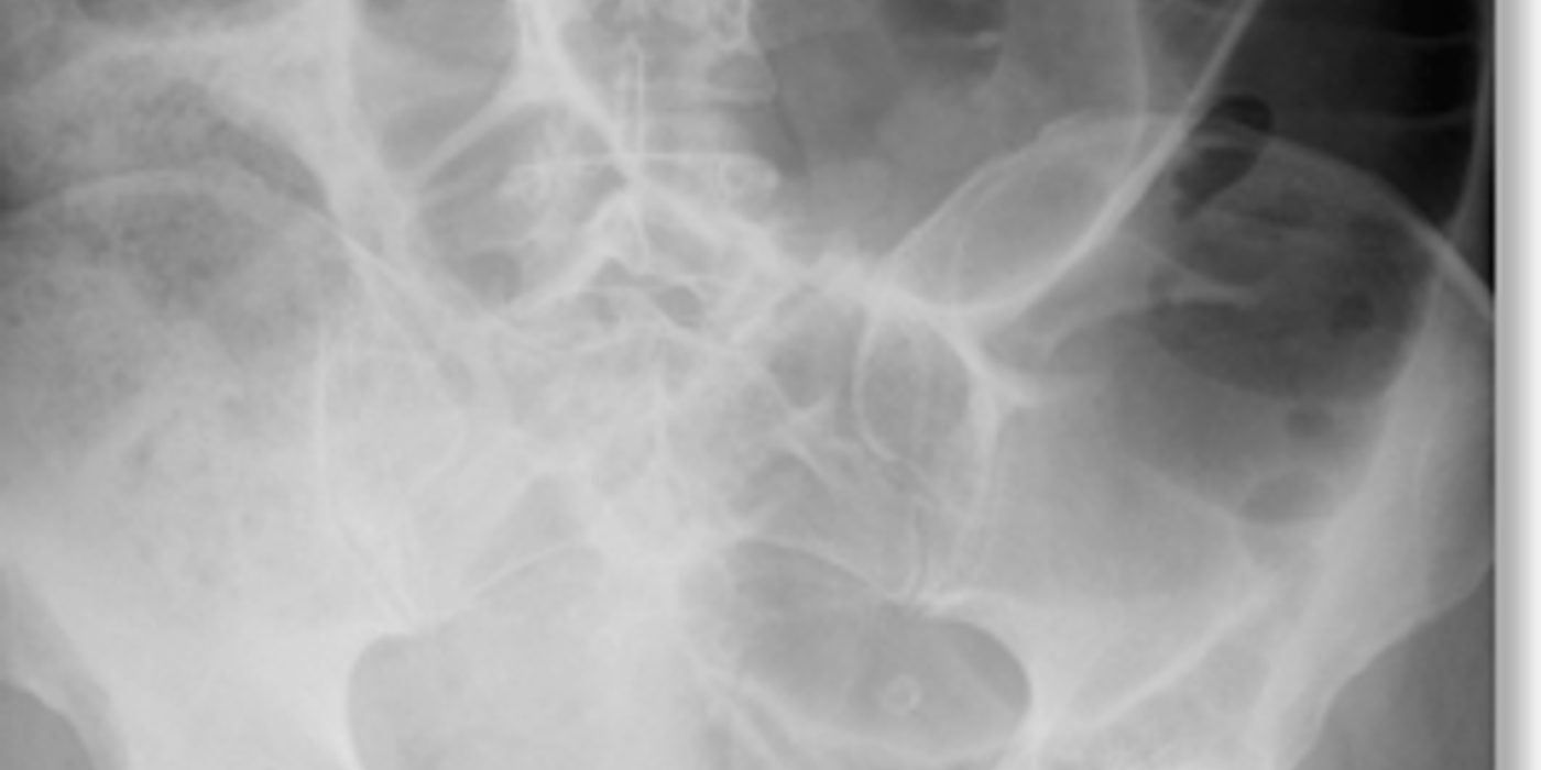
Large bowel obstruction is a potentially life-threatening blockage of the colon that commonly causes abdominal pain, progressive distension, constipation, and reduced passage of gas. Urgent assessment with resuscitation and contrast-enhanced computed tomography is usually needed to identify the site, severity, and cause, such as colorectal cancer, volvulus, diverticulitis, or faecal impaction.
Key insight: A patient with large-bowel obstruction, worsening abdominal pain, peritonism, shock, acidosis, or rising lactate may have ischaemia or perforation and requires urgent surgical review.
This 2026 overview explains the symptoms, causes, imaging approach, emergency management, and treatment options for large bowel obstruction.
Key takeaways
- Colorectal carcinoma is the commonest cause, then volvulus and diverticular stricture.
- A caecum beyond 9 cm, or any other colonic segment beyond 6 cm, is the plain-film threshold for concern.
- A closed loop obstruction with a competent ileocaecal valve risks caecal perforation.
- Resuscitate, decompress and image before deciding on the operation.
- Sigmoid volvulus is usually decompressed endoscopically first; caecal volvulus needs surgery.
What is large bowel obstruction?
Large bowel obstruction is impaired or complete passage of bowel contents through the colon. The blockage may affect the caecum, ascending colon, transverse colon, descending colon, or rectosigmoid region. In contrast, small bowel obstruction affects the jejunum or ileum and often causes earlier, more prominent vomiting.
The colon absorbs water and stores faeces. When its lumen becomes blocked, gas and fluid accumulate proximally. This produces progressive colonic dilatation, abdominal distension, pain, constipation, and reduced passage of flatus. Vomiting is usually a late feature because the colon is further along the gastrointestinal tract.
Large-bowel obstruction is the same clinical condition often abbreviated as LBO. LBO may be mechanical, functional, partial, or complete. Bowel obstructions differ according to their location, the speed of onset, and whether the intestine retains an adequate blood supply.
The term intestinal obstruction describes impaired movement through the intestine. Mechanical LBO has a physical transition point, whereas functional LBO has impaired motility without a fixed lesion. These distinctions guide imaging, monitoring, and treatment.
Causes and mechanisms
Mechanical obstruction results from a physical barrier. Colorectal carcinoma is the commonest cause, followed by volvulus and diverticular stricture. Cancer may narrow the lumen gradually or cause an acute blockage.
Other mechanical causes include:
- Sigmoid or caecal volvulus
- Diverticular disease causing a fibrotic stricture
- Faecal impaction
- Hernia
- Intussusception
- External compression or an inflammatory mass
A volvulus occurs when a mobile segment twists around its mesentery. This causes both obstruction and possible vascular compromise. In a closed-loop obstruction, a competent ileocaecal valve prevents decompression into the small bowel. Pressure can then rise rapidly, especially in the caecum. On a plain abdominal radiograph, caecal dilatation beyond 9 cm, or colonic dilatation beyond 6 cm elsewhere, raises concern for complications.
A colonic volvulus is twisting of the colon around its mesentery. Sigmoid volvulus is the most frequent form, but caecal volvulus and other volvulus patterns can produce LBO. Torsion may obstruct venous drainage first, followed by arterial compromise, ischaemia, necrosis, and perforation.
Caecal volvulus involves abnormal movement and rotation of the caecum and ascending colon. A caecal volvulus may create a closed-loop obstruction, while a sigmoid volvulus usually affects the sigmoid colon. Both conditions can progress quickly when torsion compromises the mesenteric blood supply.
Functional obstruction occurs without a physical blockage. Ileus causes reduced intestinal propulsion, often after surgery, infection, electrolyte disturbance, or medication use. Acute colonic pseudo-obstruction, also called Ogilvie syndrome, causes marked colonic dilatation without a mechanical lesion. Toxic megacolon is different: it is severe colitis with systemic toxicity and colonic dilatation. It requires urgent treatment because perforation can develop.
Ogilvie syndrome is acute colonic pseudo-obstruction without a fixed transition point. Unlike mechanical LBO, Ogilvie syndrome may improve after correcting triggers, using neostigmine, or performing colonoscopic decompression. Pseudo-obstruction, ileus, and toxic megacolon are separate diagnoses despite overlapping symptoms.
Toxic megacolon is severe colonic inflammation with systemic toxicity and marked dilatation. Toxic megacolon may complicate ulcerative colitis, infectious colitis, or other inflammatory conditions. Unlike uncomplicated pseudo-obstruction, toxic megacolon requires urgent assessment for sepsis, ischaemia, and perforation.
Untreated obstruction compromises venous drainage and arterial blood flow. The bowel may become ischaemic, necrotic, and perforated, leading to peritonitis and sepsis. Fluid sequestration, vomiting, and poor intake can also cause dehydration, renal impairment, and electrolyte disturbance. This makes the condition a surgical emergency requiring early assessment and resuscitation.
In LBO, the intestine becomes progressively dilated upstream from the blockage. The caecum is particularly vulnerable because it may have the largest diameter and highest wall tension. A closed-loop LBO can therefore develop ischaemia even before obvious free perforation appears.
Why is large bowel obstruction dangerous? Rising intraluminal pressure can cause bowel ischaemia, caecal perforation, peritonitis, sepsis, and severe fluid or electrolyte imbalance.
Large bowel obstruction is usually mechanical, commonly caused by colorectal cancer, volvulus, or diverticular stricture, and can rapidly progress to ischaemia, perforation, and sepsis.
What Symptoms and Signs Suggest Colonic Obstruction?
A large bowel obstruction usually develops more slowly than small bowel obstruction, as described in discussions of subacute and chronic intestinal obstruction and its diagnosis. The clinical picture depends on the cause, location, and whether the obstruction is complete.
LBO symptoms usually reflect accumulation of gas, stool, and fluid within the intestine. The most important symptoms are abdominal pain, abdominal distension, constipation, obstipation, nausea, and late vomiting. Partial bowel obstructions may still allow some stool or flatus to pass.
- Progressive abdominal distension is common, especially when the blockage lies in the distal colon or rectum.
- Colicky abdominal pain reflects bowel contractions attempting to push contents beyond the obstruction; constant pain suggests ischaemia or perforation.
- Constipation may progress to obstipation, meaning complete failure to pass stool or flatus through the obstructed bowel.
- Nausea and vomiting can occur, but vomiting usually develops later than with small bowel obstruction.
- Altered bowel habit, rectal bleeding, weight loss, and anaemia should raise suspicion of colorectal cancer causing colonic obstruction.
Colorectal carcinoma is the commonest cause, followed by volvulus and diverticular stricture.
Abdominal pain may be intermittent when the colon contracts against a narrowing. Persistent abdominal pain, guarding, fever, tachycardia, or acidosis increases concern for ischaemia. Faecal impaction can mimic LBO, particularly in older or immobile adults, but impaction does not exclude a more proximal obstructing lesion.
Examination findings and red flags
The abdomen may be visibly distended, with prominent or visible peristalsis. Percussion often produces a tympanic or hyperresonant note. Early bowel sounds may be high-pitched or “tinkling”; later, they can become quiet. Tenderness requires careful assessment, particularly if it is localised or severe.
A digital rectal examination may find an empty rectum, although a low tumour or impacted stool can alter this finding. Assess hydration carefully. Dry mucous membranes, tachycardia, hypotension, reduced urine output, and lethargy suggest significant fluid loss.
- Severe tenderness, guarding, rebound pain, fever, tachycardia, or rising lactate suggest strangulation, bowel ischaemia, or perforation.
A competent ileocaecal valve can create a closed-loop obstruction. Pressure then rises within the caecum, increasing perforation risk. On plain radiography, a caecum above 9 cm, or other colonic segments above 6 cm, suggests dangerous dilatation. Perforation may cause free air, peritonitis, and systemic toxicity.
- Toxic megacolon presents with marked colonic dilatation, systemic toxicity, fever, tachycardia, abdominal tenderness, and often bloody diarrhoea.
Distension, absolute constipation, late vomiting, an empty rectum, and peritoneal signs point toward serious colonic obstruction.
What causes large bowel obstruction?
The problem is a blocked colon that prevents stool and gas from passing. Causes may arise inside the lumen, within the bowel wall, or outside the bowel. The obstruction may be mechanical or functional. Cancer, volvulus, and diverticular strictures are high-yield MRCS causes.
LBO may result from colorectal cancer, diverticulitis, inflammatory strictures, faecal impaction, volvulus, intussusception, or external compression. The descending colon and sigmoid colon are common sites because their lumen is relatively narrow and stool is more solid.
The direct answer is that colorectal carcinoma is the commonest cause of large bowel obstruction. Tumours most often obstruct in the sigmoid or descending colon. These segments have narrower lumens and firmer stool. A mass may cause gradual constipation or sudden complete obstruction.
Diverticulitis can produce mural oedema, inflammation, or a fibrotic stricture. A stricture may be short and benign or may resemble colorectal cancer on symptoms and imaging. Ulcerative colitis can also cause chronic strictures and toxic megacolon. These bowel obstructions require evaluation of both the lumen and the intestinal wall.
Volvulus: sigmoid versus caecal
Sigmoid volvulus occurs when a mobile, elongated sigmoid colon twists around its mesentery. The anatomy and configuration of this segment are discussed in this overview of the sigmoid colon. It typically affects older adults with chronic constipation, reduced mobility, or neurological disease. Abdominal radiography may show a large “coffee-bean” loop. CT can demonstrate a twisted mesenteric pedicle, known as the whirl sign. Ischaemia and perforation become more likely if decompression fails.
A sigmoid colon volvulus is a form of colonic volvulus caused by torsion of a mobile sigmoid loop. The splenic flexure, descending colon, and sigmoid colon are less mobile than the caecum, but anatomical variation can permit twisting. CT and plain abdominal radiograph findings help distinguish sigmoid volvulus from other bowel obstructions.
Caecal volvulus results from abnormal caecal mobility or incomplete fixation. Patients are often younger than those with sigmoid volvulus. The caecum may move towards the upper abdomen or left side. Imaging can show a displaced, dilated caecum and a whirl sign. Unlike uncomplicated sigmoid volvulus, endoscopic decompression is less reliable. Early surgery is usually required because bowel compromise can develop rapidly.
Caecal volvulus can displace the caecum toward the left upper abdomen, where it may be confused with a dilated transverse or descending colon. A caecal transition point and mesenteric swirl on computed tomography support the diagnosis. Repeated episodes of caecal volvulus can cause recurrent LBO and progressive intestinal injury.
A competent ileocaecal valve creates a closed-loop obstruction. This prevents decompression into the small bowel and raises pressure within the caecum. On a plain film, caecal dilatation beyond 9 cm, or colonic dilatation beyond 6 cm elsewhere, increases concern for perforation.
Benign and functional causes
Diverticular disease can produce a fibrotic sigmoid stricture, while inflammation from diverticulitis can lead to complications affecting the bowel wall and surrounding tissues (What causes diverticulitis?). Other benign strictures result from Crohn’s disease, ulcerative colitis, previous colorectal surgery, anastomosis, or pelvic radiotherapy. Adhesions and external compression are less common causes in the large bowel than in the small bowel.
Diverticulitis may narrow the sigmoid colon through inflammation and scarring. A diverticulitis-related stricture can produce partial bowel obstruction, while active diverticulitis may cause abscess, sepsis, or perforation. Ulcerative colitis increases the risk of inflammatory strictures and toxic megacolon.
The differential diagnosis also includes faecal impaction, foreign material, and intussusception. Gallstone ileus usually causes small bowel obstruction, but a gallstone can rarely enter the colon through a biliary-colonic fistula. Acute colonic pseudo-obstruction causes severe dilation without a physical blockage. Toxic megacolon causes systemic toxicity and colonic dilation, usually during severe inflammatory or infective colitis.
Faecal impaction is retained, hardened stool that prevents normal evacuation. Severe faecal impaction may cause abdominal pain, overflow diarrhoea, rectal pressure, and apparent LBO. Digital examination may identify distal impaction, but computed tomography is appropriate when symptoms are severe, atypical, or associated with distension.
Identify the cause and site, then assess for closed-loop obstruction, ischaemia, and perforation.
How Is Large Bowel Obstruction Diagnosed?
Diagnosis combines urgent clinical assessment, blood tests, and imaging. CT abdomen and pelvis with contrast usually identifies the obstruction’s level, cause, and complications in a stable patient.
The diagnostic imaging approach for LBO should answer four questions: where is the blockage, what produced it, is the bowel viable, and has a complication developed? Imaging may include a plain abdominal radiograph, abdominal radiograph with chest imaging, ultrasound in selected patients, and contrast-enhanced computed tomography.
Initial clinical assessment
Start with an ABCDE assessment. Check the airway, breathing, circulation, disability, and exposure. Look for shock, dehydration, hypoxia, confusion, or signs of sepsis. Assess temperature, heart rate, blood pressure, respiratory rate, oxygen saturations, and urine output.
Ask about pain, distension, vomiting, constipation, and the last passage of stool or flatus. Establish the onset and progression of symptoms. Ask about previous abdominal surgery, diverticular disease, colorectal cancer, hernias, and previous episodes of volvulus.
Examine the abdomen for distension, scars, visible peristalsis, tenderness, guarding, and peritonism. Listen for high-pitched or absent bowel sounds, although bowel sounds cannot confirm or exclude obstruction. Palpate for masses, including a possible colonic tumour.
Perform a digital rectal examination. An empty rectum supports distal obstruction, but does not prove it. Check for rectal masses, blood, faeces, and pain. Look for sepsis or peritonitis, which may indicate bowel ischaemia or perforation.
Blood tests
Blood tests assess physiological disturbance and prepare the patient for treatment or surgery. Request:
- Full blood count for anaemia, infection, and leucocytosis.
- Urea and electrolytes to identify dehydration, renal injury, and electrolyte imbalance.
- C-reactive protein or other inflammatory markers for inflammation.
- Venous or arterial blood gas, including lactate, to assess tissue hypoperfusion.
- Clotting studies before invasive procedures or surgery.
- Group and save, particularly when an operation may be required.
- Blood cultures when the patient has fever, rigors, or suspected sepsis.
A raised lactate can suggest bowel ischaemia, but a normal result does not exclude early strangulation. Clinical deterioration requires urgent senior and surgical review.
Imaging and endoscopy
An abdominal radiograph may show dilated colon with haustral markings. A caecal diameter above 9 cm, or another colonic diameter above 6 cm, is concerning for significant dilatation. An erect chest radiograph can identify free subdiaphragmatic gas from perforation.
A plain abdominal radiograph is quick and may show a coffee-bean sign, a displaced caecum, or diffuse megacolon. However, an abdominal radiograph cannot reliably identify the underlying lesion, early ischaemia, or a small perforation.
CT abdomen and pelvis with intravenous contrast is usually the preferred test for a stable patient. It can locate the transition point, identify an obstructing colonic lesion, and detect volvulus, ischaemia, perforation, or metastatic disease.
Contrast-enhanced computed tomography provides cross-sectional assessment of the entire abdomen and pelvis. Contrast-enhanced computed tomography images may show a tumour, diverticular stricture, caecal volvulus, sigmoid volvulus, ischaemia, free air, or abscess.
Modern computed tomography and multidetector CT, or MDCT, provide rapid visualisation of the intestine. An MDCT scan can reconstruct the colon in multiple planes. Computed tomography images may show a transition point that is not visible on a plain abdominal radiograph.
A second contrast-enhanced computed tomography examination may be considered when the patient deteriorates or the initial scan was limited. In patients with renal impairment or contrast allergy, the radiologist can adapt the imaging protocol. The choice between CT, MDCT, and noncontrast tomography depends on stability and diagnostic needs.
A sigmoid volvulus may produce the coffee-bean sign, caused by a massively dilated loop. Caecal volvulus may show a displaced, dilated caecum. CT can also demonstrate a twisted mesentery and the precise site of obstruction.
On contrast-enhanced computed tomography images, reduced bowel-wall enhancement, mesenteric oedema, pneumatosis, portal venous gas, or free fluid may suggest ischaemia. Computed tomography images can also distinguish megacolon, pseudo-obstruction, and mechanical LBO when the transition point is unclear.
A water-soluble contrast enema can define the distal colon when CT is unavailable or when sigmoid volvulus is suspected. It may show a tapering “bird’s-beak” appearance. Endoscopy can confirm and sometimes decompress sigmoid volvulus, but suspected perforation or peritonitis requires urgent surgical management.
What is the key diagnostic test? CT with contrast is generally the key test because it identifies the cause, level, and complications of bowel obstruction. Diagnose suspected large bowel obstruction with rapid resuscitation, focused examination, targeted blood tests, and timely CT imaging.
How do you resuscitate and initially manage it?
LBO management begins with resuscitation, bowel rest, correction of physiological abnormalities, and early surgical consultation. Multidisciplinary decision-making commonly includes emergency physicians, surgeons, radiologists, gastroenterologists, anaesthetists, and critical-care clinicians.
What are the immediate priorities?
What: Admit the patient under the on-call surgical team and treat suspected large bowel obstruction as a potentially urgent condition. Keep the patient nil by mouth (NBM) and insert two large-bore intravenous cannulas.
Why: Vomiting, third-spacing, and reduced oral intake cause dehydration and electrolyte loss. A distended bowel also increases aspiration and perforation risks. Early resuscitation creates a safer pathway to endoscopy or surgery.
How: Begin intravenous crystalloid fluids, guided by blood pressure, heart rate, peripheral perfusion, lactate, and comorbidities. Check and correct sodium, potassium, urea, creatinine, magnesium, and acid–base abnormalities. Give suitable analgesia and antiemetics, while avoiding excessive sedation that could mask deterioration.
Insert a urinary catheter when the patient is significantly unwell, undergoing active resuscitation, or likely to need surgery. Record hourly urine output. A practical target is approximately 0.5 mL/kg/hour in adults, alongside improving clinical observations.
Repeat abdominal examinations and monitor pulse, blood pressure, respiratory rate, oxygen saturation, temperature, pain, abdominal distension, fluid balance, and lactate. Resuscitation should continue while investigations and operative planning occur. Initial treatment includes fluid replacement, electrolyte correction, aspiration-risk reduction, and gastrointestinal decompression.
What is the role of decompression and escalation?
What: Use a nasogastric tube if there is persistent vomiting, marked proximal bowel dilatation, or a high aspiration risk. Its role is more limited than in small bowel obstruction because the obstruction lies further downstream.
Why: A nasogastric tube mainly decompresses the stomach and proximal small bowel. It may help when the ileocaecal valve is incompetent, allowing colonic contents and gas to reflux proximally. It does not reliably decompress a closed distal colon or sigmoid volvulus.
How: Consider broad-spectrum intravenous antibiotics when there is suspected perforation, peritonitis, bowel ischaemia, sepsis, or planned emergency surgery. Discuss the patient early with anaesthetics. Involve critical care for shock, rising lactate, organ dysfunction, severe electrolyte disturbance, or escalating oxygen and fluid requirements.
Prepare for urgent operation when there is peritonitis, free perforation, suspected ischaemia, closed-loop obstruction, clinical deterioration, or failure of conservative treatment. A competent ileocaecal valve can create a closed loop and rapidly increase caecal pressure. A caecal diameter above 9 cm, or above 6 cm elsewhere in the colon, raises concern for dangerous dilatation on plain radiography.
Urgent senior review is required for worsening pain, tachycardia, fever, hypotension, acidosis, guarding, or increasing distension. Antibiotics and resuscitation must not delay source control when perforation is likely.
Resuscitate early, decompress selectively, monitor closely, and involve senior surgical, anaesthetic, and critical-care teams before deterioration forces emergency intervention.
How does treatment differ by cause?
Large bowel obstruction is treated by relieving the blockage, correcting physiological derangement, and addressing its underlying cause. Management depends on the obstruction’s site, severity, bowel viability, contamination, and the patient’s operative risk.
Initial treatment usually includes nil by mouth, intravenous fluids, electrolyte correction, analgesia, and close monitoring. A urinary catheter can assess renal perfusion and fluid balance. Nasogastric drainage may help if vomiting or proximal distension is present. Early antibiotic therapy is appropriate when ischaemia, perforation, or contamination is suspected.
Volvulus
A suitable sigmoid volvulus without peritonitis or suspected perforation can often undergo flexible sigmoidoscopic detorsion. A flatus tube may remain temporarily to maintain decompression. Endoscopic treatment relieves the immediate obstruction but does not correct the risk of recurrence.
Definitive treatment is usually sigmoid colectomy during the same admission or soon afterwards. The surgeon may perform a primary anastomosis if the bowel is viable and the patient is stable. Hartmann’s procedure or another diverting operation may be safer when contamination, poor tissue quality, or physiological instability is present.
Caecal volvulus generally requires surgery, because endoscopic decompression is unreliable and recurrence is likely. Viable bowel may be treated with right hemicolectomy and anastomosis. Ischaemic or perforated bowel requires resection, with anastomosis or stoma formation based on patient fitness and contamination.
Malignant obstruction
Colonic stenting can decompress a malignant blockage. In selected patients, it acts as a bridge to planned surgery, allowing resuscitation and staging. It can also provide palliation when curative resection is unsuitable. Stenting is less appropriate when perforation is suspected or the anatomy is unsuitable.
Symptoms of operative choices include tumour resection, diversion with a loop stoma, or resection with primary anastomosis. The decision depends on tumour location, proximal dilatation, bowel viability, contamination, nutritional state, and the patient’s physiological reserve. A stoma may reduce anastomotic risk in an unstable or contaminated field.
Functional obstruction and faecal loading
Toxic megacolon requires urgent medical and surgical assessment. Treatment includes bowel rest, intravenous fluids, correction of electrolytes, antibiotics, and therapy for the underlying inflammatory or infective disease. Colonoscopy is usually avoided because of perforation risk. Deterioration, perforation, uncontrolled bleeding, or failure of medical treatment may require emergency subtotal colectomy.
Acute colonic pseudo-obstruction, or Ogilvie syndrome, has no mechanical blockage. Supportive care and treatment of triggers may be sufficient. Persistent or severe cases may respond to intravenous neostigmine, provided contraindications are excluded. Colonoscopic decompression is an alternative, while perforation or refractory distension may require surgery.
Non-perforated faecal impaction may respond to digital disimpaction, enemas, or both; severe constipation can become life-threatening when diagnosis or treatment is delayed, as illustrated by a reported constipation-related death (Sally Lewis: New date set for inquest after Bromsgrove constipation death). A maintenance bowel regimen helps prevent recurrence. This approach must not delay imaging or surgery when peritonitis, ischaemia, or complete obstruction is suspected.
Faecal impaction treatment depends on the location and severity of the impaction. Options include suppositories, enemas, manual disimpaction, and oral osmotic laxatives after a dangerous obstruction has been excluded. Recurrent impaction requires review of hydration, mobility, medications, diet, and pelvic-floor function.
Perforation requires urgent resuscitation, broad-spectrum antibiotics, source control, and emergency operation. The procedure may involve resection, anastomosis, or diversion, depending on contamination and physiological stability.
The cause and condition of the bowel determine treatment: decompress suitable sigmoid volvulus, operate early for caecal volvulus or perforation, and tailor cancer surgery or diversion to patient risk.
This quick-reference table summarises large bowel obstruction treatment according to the clinical situation, highlighting initial supportive care, decompression for suitable sigmoid volvulus, and escalation when bowel ischaemia, perforation, or contamination is suspected.
What does the examiner want on large bowel obstruction?
Core revision points
- Large bowel obstruction is usually mechanical, with colorectal carcinoma the commonest cause, followed by volvulus and diverticular stricture.
- Mechanical obstruction blocks intestinal contents at a physical point; volvulus adds twisting, closed-loop obstruction, and threatened vascular compromise.
- Acute colonic pseudo-obstruction causes severe colonic dilatation without a physical blockage, often affecting seriously ill or postoperative patients.
- Ileus produces diffuse bowel inactivity, usually after surgery, inflammation, metabolic disturbance, or medication, without a transition point.
- Toxic megacolon combines systemic toxicity, colitis, and colonic dilatation; fever, tachycardia, shock, or peritonitis demand urgent escalation.
- Sigmoid volvulus typically causes marked distension and a coffee-bean sign; initial treatment may involve endoscopic decompression if stable.
- Caecal volvulus often produces small-bowel obstruction features, with a displaced caecum and higher risk of rapid strangulation.
- Colorectal cancer usually causes progressive, asymmetrical narrowing, altered bowel habit, weight loss, and obstruction near the tumour.
- Plain-film thresholds are caecal diameter above 9 cm or other colonic diameter above 6 cm, indicating significant dilatation risk.
- A competent ileocaecal valve creates a closed loop, raising caecal pressure and risking perforation; perforation causes peritonitis and requires urgent surgery.
- Never let imaging delay resuscitation: keep the patient nil by mouth, give intravenous fluids, monitor urine output, correct electrolytes, and seek senior help.
- Examine systematically using inspection, auscultation, percussion, and palpation; document distension, tenderness, bowel sounds, hernias, masses, and rectal findings.
- The MRCS applied surgical science and critical care station awards 12 marks for knowledge, 4 for skills, and 4 for professionalism.
- Practise this topic through Mrcspartbquestions interactive scenarios, anatomy spot tests, imaging questions, explanations, mark schemes, and cross-device progress tracking.
What is the key surgical red flag? Peritonitis, perforation, strangulation, systemic deterioration, or failed decompression requires urgent operative assessment.
Recognise the obstruction pattern, resuscitate early, identify threatened bowel, and escalate promptly when volvulus or peritonitis is present.
How is this topic marked in MRCS Part B?
MRCS Part B marks an applied surgical science and critical care station out of 20: 12 marks for clinical knowledge and its application, 4 for clinical and technical skill and 4 for professionalism. Three of the seventeen examined stations fall in this area.