MRCS Part B Knowledge Library
Topic-by-topic guides mapped to the MRCS Part B OSCE — anatomy, applied surgical sciences and the reasoning each station expects.
Applied Surgical Sciences
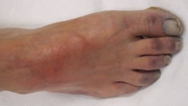
Acute Limb Ischaemia for MRCS Part B: The Six Ps and Management
Acute limb ischaemia is a sudden fall in arterial blood flow that threatens limb viability and demands urgent assessment, anticoagulation and, when indicated, revascularisation. It typically develops within 14 days and may result from embolism, in-situ thrombosis or acute-on-chronic disease. Pain, pallor, pulselessness, paraesthesia, paralysis and poikilothermia signal increasing severity and possible irreversible injury.
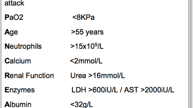
Acute Pancreatitis for MRCS Part B: Causes, Glasgow Criteria and Management
Acute pancreatitis is sudden inflammation of the pancreas caused by premature activation of digestive enzymes, often linked to gallstones or alcohol. It typically presents with severe upper abdominal pain, nausea, and vomiting, and may lead to pancreatic necrosis, respiratory failure, or shock. Diagnosis and severity assessment rely on clinical findings, serum lipase, and targeted imaging.
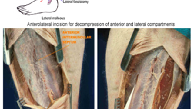
Compartment Syndrome for MRCS Part B: Signs, Pressures and Fasciotomy
Spot compartment syndrome early by recognising severe, progressive pain—especially pain with passive stretching—alongside a tense, swollen limb, tingling, or weakness. It is caused by rising pressure within a closed muscle compartment, restricting blood flow to nerves and muscles. Acute cases can cause permanent damage within hours, and a normal distal pulse does not rule it out.
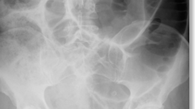
Large Bowel Obstruction for MRCS Part B: Causes, Imaging and Management
Large bowel obstruction causes progressive abdominal pain, distension, constipation, and reduced passage of flatus; vomiting is usually late. Treatment involves urgent assessment, bowel rest, intravenous fluids, electrolyte correction, and decompression, followed by endoscopic, stent, or surgical management according to the cause. Colorectal carcinoma is the commonest cause, followed by volvulus and diverticular stricture.
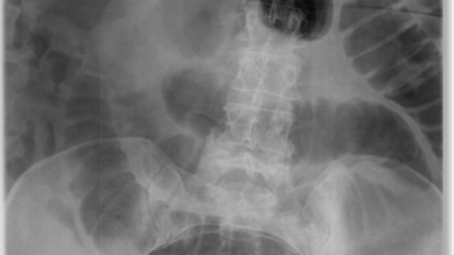
Small Bowel Obstruction for MRCS Part B: Causes, Imaging and Management
Small bowel obstruction is treated initially with bowel rest, nasogastric decompression, intravenous fluids and electrolyte correction, with urgent surgery for strangulation, perforation, peritonitis, or failure to resolve. Adhesions from previous abdominal surgery cause approximately 65–75% of cases in developed countries, so management also focuses on identifying the obstruction’s cause and complications.
Clinical Examination
Abdominal Examination for MRCS Part B: Inspection, Palpation and the Organs
Examine the abdomen flat with one pillow, exposed from nipples to knees in practice covered to the groin: inspect, palpate lightly then deeply, examine liver, spleen and kidneys, percuss for ascites and auscultate.
Breast Examination for MRCS Part B: Positions, Quadrants and Triple Assessment
Inspect the breasts in four positions, palpate the normal side first through all four quadrants and the axillary tail, examine the nipple and the regional nodes, then offer the components of triple assessment.
Cardiovascular Examination for MRCS Part B: Sequence, Signs and Presentation
Examine the cardiovascular system in a fixed order: end of the bed, hands, pulse, blood pressure, JVP, face, praecordium, then auscultation in four areas with a finger on the carotid, finishing with peripheral pulses and oedema.
Cranial Nerve Examination for MRCS Part B: Twelve Nerves in Order
Test the cranial nerves in numerical order: smell, vision and pupils, eye movements, facial sensation and the muscles of mastication, facial movement, hearing, palate and gag, accessory muscles and the tongue.
Elbow Examination: A Verified Sequence for the MRCS Part B Station
Examine the elbow by looking at the carrying angle and skin, palpating the epicondyles and olecranon, testing flexion, extension, pronation and supination, then adding resisted tests for tennis elbow and Tinel's sign over the ulnar nerve.
Foot and Ankle Examination: Alignment, Movement, Achilles and the Ottawa Rules
Examine the foot and ankle standing and lying, working from hindfoot to forefoot, then test movement, the Achilles tendon with the calf squeeze, and apply the Ottawa ankle and foot rules after injury.
Hand and Wrist Examination: Look, Feel, Move, Function and the Nerves
Examine the hand palms down then palms up, palpate the wrist and small joints, test movement, then finish with function and the median, ulnar and radial nerves; NICE CKS warns that Phalen's and Tinel's tests must not be used in isolation.
Hernia Examination for MRCS Part B: Standing, Supine and the Cough Impulse
Examine a groin hernia standing first, then supine: inspect, ask the patient to cough, palpate the lump, decide whether you can get above it and whether it is reducible, and locate it against the pubic tubercle.
Hip Examination: Gait, Trendelenburg, Fixed Flexion and Rotation
Start the hip examination with gait, then look, feel and move, testing internal rotation with the hip flexed because NICE CKS identifies painful restricted internal rotation as the classic sign of hip osteoarthritis.
Knee Examination: Effusion, Ligaments, Menisci and the Ottawa Knee Rule
Examine the knee with look, feel, move and stability testing; NICE CKS gives a normal range of zero to 135 degrees, supports Lachman and drawer testing, and states that meniscal tests such as McMurray's have particularly poor diagnostic accuracy.
Lower Limb Neurological Examination: Tone, Power, Reflexes, Sensation and Gait
Run the lower limb neurological examination in the standard order of inspection, tone, power, reflexes, coordination, sensation and gait, grading power on the published 0 to 5 scale and testing the ISNCSCI key muscles.
Lumps and Bumps Examination for MRCS Part B: A System for Any Lump
Examine any lump with the same system: site, size, shape, surface, edge, colour, temperature, tenderness, consistency, fluctuance, transillumination, pulsatility, compressibility, fixity and the regional lymph nodes.
Per Rectum (PR) Examination for MRCS Part B: Consent, Technique and Findings
A rectal examination is an intimate examination: explain it, gain consent, offer a chaperone, position the patient in the left lateral position, inspect the perianal skin, then assess tone, the rectal wall and the prostate.
Peripheral Vascular Examination for MRCS Part B: Pulses, ABPI and Buerger's Test
Examine both legs together: inspect for tissue loss and colour change, assess temperature, capillary refill and sensation, palpate every pulse from the aorta down, then perform Buerger's test and measure the ABPI.
Respiratory Examination for MRCS Part B: Inspection to Auscultation
The respiratory examination is inspection, palpation for expansion and trachea, percussion and auscultation, repeated front and back, with the percussion note and breath sounds interpreted together to name the pathology.
Shoulder Examination: Look, Feel, Move and the Special Tests That Count
Examine the shoulder from front, side and behind, palpate the bony landmarks, then test active, passive and resisted movement before any special test; NICE CKS puts the painful arc between 70 and 120 degrees of active abduction.
Spine and Neurological Examination: Movement, Root Signs and Cauda Equina
Examine the spine by region with look, feel and move, then screen the limbs neurologically; a straight leg raise is positive when pain is reproduced below 60 degrees, and cauda equina symptoms need emergency referral for MRI.
Testicular Examination for MRCS Part B: Scrotal Lumps and What They Mean
Examine the scrotum standing then supine: inspect, palpate both testes and epididymes, ask whether you can get above the lump, test for a cough impulse, transilluminate, and examine the groin nodes and abdomen.
Thyroid Examination for MRCS Part B: Neck Lumps and Thyroid Status
Examine the thyroid from the front and then from behind: inspect the neck with the patient swallowing and protruding the tongue, palpate both lobes and the nodes, percuss the sternum and auscultate for a bruit.
Upper Limb Neurological Examination for MRCS Part B: Tone, Power, Reflexes and Sensation
Examine the upper limbs in a fixed order: inspection and pronator drift, tone, power graded on the MRC scale, the biceps, triceps and supinator reflexes, sensation by dermatome and modality, then coordination.
Varicose Vein Examination for MRCS Part B: Distribution, Skin Changes and Special Tests
Examine varicose veins with the patient standing: map the long and short saphenous distributions, look for skin changes of chronic venous hypertension, palpate for a saphena varix, then perform the tap, tourniquet and Trendelenburg tests.
Procedural Skills
Cannulation: Aseptic Insertion, Flushing and Setting Up an Infusion
Cannulation is marked on aseptic non-touch technique and safety: decontaminate the skin with chlorhexidine gluconate in 70% alcohol, advance the cannula rather than the needle, flush with sterile sodium chloride, and remove the device as soon as it is no longer needed.
Central Venous Catheter Insertion: Ultrasound, Seldinger Technique and Never Events
NICE TA49 recommends two-dimensional ultrasound guidance as the preferred method for internal jugular central venous catheter insertion in elective cases, and a retained guidewire is a Never Event, so the wire is held and counted at every step.
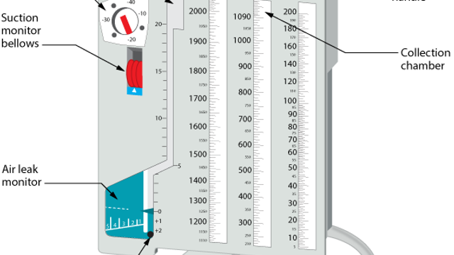
Chest Drain Insertion for MRCS Part B: Triangle of Safety, Technique and Marking
Chest drain insertion involves assessing the indication and ABCDE stability, obtaining consent, preparing sterile equipment, positioning the patient, anaesthetising the site, and advancing a tube into the pleural space to drain air, blood, pus, or fluid. This guide covers drain function, suction, complications, and MRCS Part B assessment points, including ultrasound for pleural effusions.
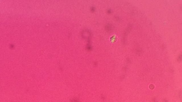
Joint Aspiration for MRCS Part B: Asepsis, Technique and Synovial Fluid
Knee joint aspiration works by inserting a sterile needle into the knee joint to withdraw excess synovial fluid for pressure relief or laboratory testing. Also called arthrocentesis, it can identify infection, bleeding, crystals, or inflammatory arthritis; fluid is typically assessed with Gram stain, culture, crystal microscopy, and cell count.
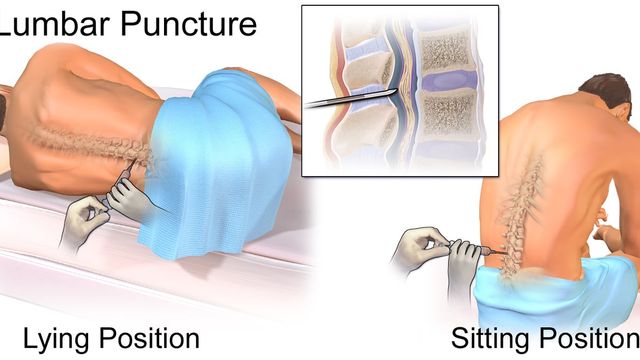
Lumbar Puncture for MRCS Part B: Landmarks, Layers and Technique
A lumbar puncture involves inserting a hollow needle into the lower back to collect cerebrospinal fluid (CSF) for testing. You are positioned on your side, and the needle enters below the spinal cord, usually at L3/L4 or L4/L5. Results can help investigate infection, inflammation, bleeding, or abnormal pressure.
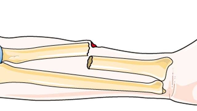
Open Fractures for MRCS Part B: Gustilo-Anderson and Initial Management
Open fractures are emergencies: cover the wound with saline-soaked gauze, avoid pushing exposed bone back, assess circulation and sensation, splint above and below the injury, and arrange urgent hospital care. Treatment typically includes intravenous broad-spectrum antibiotics, tetanus prophylaxis, surgical debridement, fracture stabilisation, and soft-tissue coverage; the Gustilo-Anderson classification guides severity after debridement.
Urethral Catheterisation: Aseptic Insertion, the Balloon and the Foreskin
Urethral catheterisation is an aseptic procedure: clean the meatus, use lubricant from a single-use container, inflate the balloon with 10 mL of sterile water in an adult only once urine drains, and replace the foreskin at the end.
Venepuncture: Aseptic Technique, Order of Draw and Blood Cultures
Venepuncture is marked on identification, asepsis and sharps safety: clean the site with 70% alcohol for 30 seconds and let it dry, never re-palpate, release the tourniquet before withdrawing the needle, and label at the bedside.
Surgical Pathology
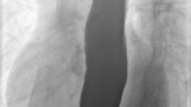
Achalasia for MRCS Part B: Pathophysiology, Diagnosis and Treatment
Achalasia is an oesophageal motility disorder in which the lower oesophageal sphincter fails to relax and peristalsis is absent or ineffective, causing progressive difficulty swallowing food and liquids. Regurgitation, chest pain, nocturnal cough, and weight loss are also common. High-resolution manometry confirms the diagnosis, while pneumatic dilation, Heller myotomy, or peroral endoscopic myotomy can improve oesophageal emptying.
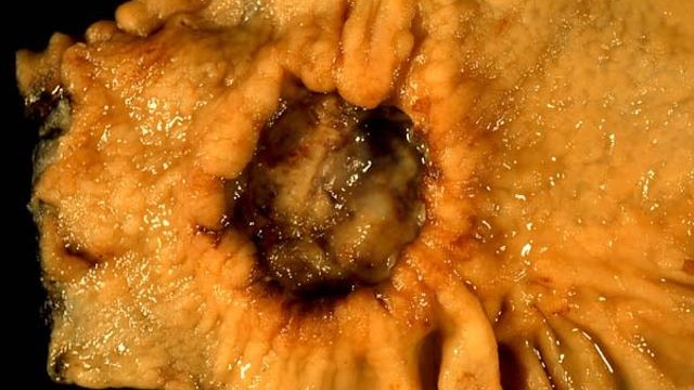
Gastric Carcinoma for MRCS Part B: Risk Factors, Spread and Staging
Gastric carcinoma is stomach cancer that may cause indigestion, weight loss, vomiting, or gastric outlet obstruction; it commonly develops through chronic *Helicobacter pylori*–associated mucosal damage and is treated according to TNM stage with surgery, systemic therapy, or palliation. Adenocarcinoma accounts for about 90% of stomach cancers, including intestinal and diffuse types such as linitis plastica.
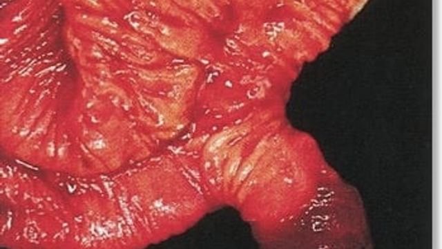
Meckel's Diverticulum for MRCS Part B: The Rule of 2s and Presentation
Meckel's diverticulum is a congenital true diverticulum of the ileum caused by incomplete obliteration of the omphalomesenteric duct. It may cause painless gastrointestinal bleeding, abdominal pain, inflammation, or bowel obstruction, while asymptomatic cases need no treatment and symptomatic cases often require surgical resection, particularly when ectopic gastric mucosa is present.

Oesophageal Carcinoma for MRCS Part B: Types, Barrett's and Staging
Oesophageal carcinoma is a cancer of the oesophageal lining that commonly causes progressive dysphagia, arises as squamous cell carcinoma or adenocarcinoma, and is treated according to stage with endoscopic therapy, chemotherapy, radiotherapy or surgery. Difficulty swallowing solids before liquids is typical; Barrett’s oesophagus is a key adenocarcinoma risk factor, and the oesophagus is approximately 25 cm long.
FAQ
What is the Knowledge library?
A free, evidence-based reference covering the orthopaedic conditions that matter for the ST3 interview and day-to-day practice — management-focused, anchored to the current BOAST and NICE standards, and reviewed against the live guideline text.
How is each article kept accurate?
Every guideline claim is checked against the current published standard (BOAST, NICE, GIRFT) and each article lists its key evidence. The last-reviewed date is shown on every article.
How does this help with the ST3 interview?
Each condition links to how it comes up at interview — the station vignette and opening questions — and the question bank turns the same topics into full station practice with model answers.