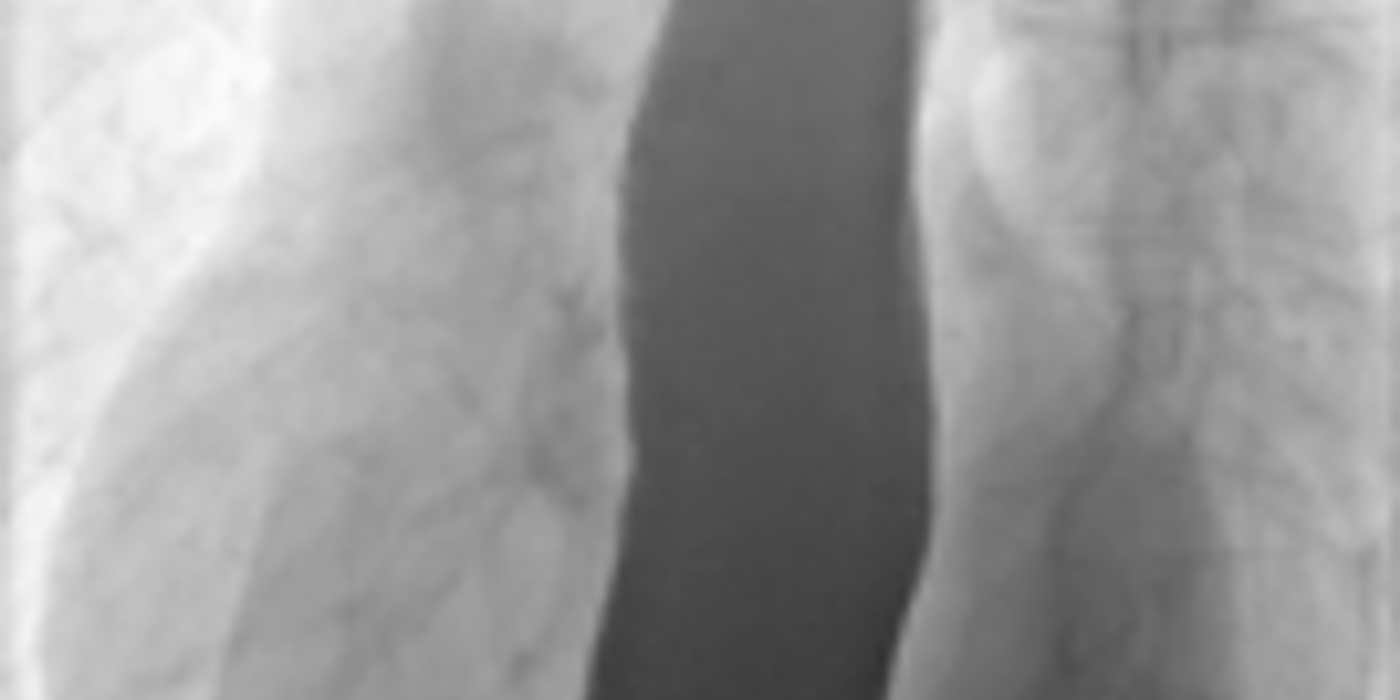
Achalasia is a rare condition in which the oesophagus cannot move food normally and the lower oesophageal sphincter fails to relax. Doctors typically confirm it with endoscopy, barium swallow, and high-resolution manometry, then use pneumatic dilation, POEM, surgery, or medication to improve passage into the stomach. Long-term follow-up is important because symptoms can recur and complications may develop.
Key takeaways
- Achalasia is failure of the lower oesophageal sphincter to relax, with loss of oesophageal peristalsis.
- The classic barium swallow shows a dilated oesophagus tapering to a bird's beak.
- Oesophageal manometry is the diagnostic standard.
- Endoscopy is needed to exclude pseudoachalasia from a tumour at the cardia.
- Treatment options are pneumatic dilatation, Heller's myotomy and peroral endoscopic myotomy.
How does achalasia present?
Achalasia symptoms often come on gradually because the oesophagus progressively loses coordinated movement. The most recognisable pattern is trouble swallowing both solids and liquids, followed by regurgitation, chest discomfort, and weight loss.
Achalasia is an oesophageal motility disorder where the lower oesophageal sphincter fails to relax, while normal peristalsis is absent or ineffective. The term cardiospasm describes the same functional obstruction at the gastro-oesophageal junction. Food and liquid therefore remain in the oesophagus instead of passing normally into the stomach.
Trouble swallowing is called dysphagia. In achalasia, the oesophagus cannot move oesophageal contents efficiently, so swallowing food or liquid may feel slow, blocked, or incomplete.
Typical symptoms and patient history
The classic presentation is slowly progressive dysphagia to both food and liquids. Patients may first struggle with solid food, then develop difficulty swallowing drinks. This pattern helps distinguish a motility disorder from a fixed mechanical obstruction, which usually affects solids first.
Common symptoms include:
- Regurgitation of undigested food or saliva
- Retrosternal chest pain or pressure
- Nocturnal cough caused by aspiration
- Weight loss from reduced intake
- Choking episodes or disturbed sleep
- Vomiting-like episodes, although the material is usually regurgitated rather than forcefully vomited
- Heartburn-like discomfort
This combination of chest pain, regurgitation, swallowing difficulty, and unexplained weight loss is also highlighted in clinical reporting on achalasia symptoms. The Mayo Clinic describes the problem as poor relaxation of the oesophagus muscles, preventing food and drink from reaching the stomach. Symptoms may worsen after meals or when lying flat. Some patients learn to stand, stretch, or drink large amounts of water to help food pass.
Chest pain and regurgitation can mimic gastro-oesophageal reflux disease. However, reflux treatment may provide little benefit. The symptoms may also resemble a mechanical stricture, oesophageal cancer, or external compression. New dysphagia, marked weight loss, bleeding, or persistent vomiting-like symptoms require prompt investigation.
Progressive difficulty with both solids and liquids suggests a movement disorder, but testing is still needed to exclude cancer, narrowing, or pseudoachalasia.
Labelled diagram: normal passage versus cardiospasm
Text NORMAL OESOPHAGUS CARDIOSPASM / ACHALASIA Food ↓ Food ↓ Peristaltic waves Dilated oesophagus ↓ ↓ Lower sphincter relaxes Tight lower sphincter ↓ ✕ Stomach receives food Food and liquid retained
The four key clinical features are dysphagia, regurgitation, chest pain, and weight loss. A symptom score used in clinical assessment rates each from 0 to 3, producing a maximum score of 12. Recognising this pattern helps guide the next diagnostic step and appropriate treatment.
Progressive dysphagia to both food and liquids, with regurgitation and chest symptoms, should raise suspicion of achalasia or cardiospasm.
What Causes Oesophageal Emptying to Fail?
Achalasia occurs when nerve cells controlling oesophageal muscle function deteriorate, preventing the lower oesophageal sphincter from opening normally. This nerve and muscle failure is what causes achalasia rather than a simple blockage inside the stomach.
The main problem is damage to the nerves controlling the oesophagus (oesophagus). In achalasia, inhibitory neurons within the myenteric plexus degenerate. These nerves normally help the lower oesophageal sphincter (LOS) relax when swallowed food reaches it.
The lower oesophageal sphincter, abbreviated LES, is the muscular valve between the oesophagus and stomach. Persistent pressure in the lower oesophageal sphincter LES prevents the oesophagus from moving material into the stomach. The terms sphincter LES and lower oesophageal sphincter les describe this same anatomical region.
As a result, the LOS remains abnormally tight. Oesophageal peristalsis also becomes absent or severely impaired. Food and liquid then collect above the gastro-oesophageal junction instead of entering the stomach. This explains progressive swallowing symptoms, regurgitation, chest discomfort, and sometimes aspiration.
When the oesophagus cannot move oesophageal contents, swallowing disorders may worsen over time. A person may initially compensate by eating slowly, drinking water, or changing posture, but the oesophagus may later become enlarged. This is why achalasia cardiospasm is considered a disorder of movement between the oesophagus and stomach, not a primary stomach disease.
Proposed causes and risk considerations
- Degeneration of myenteric inhibitory neurons prevents lower oesophageal sphincter relaxation and disrupts normal oesophageal peristalsis.
- The exact cause remains unknown, although autoimmune inflammation, infections, and inherited genetic factors have all been investigated. Emerging genetic-risk research is also examining the possible immune basis of achalasia.
- The condition is sometimes called cardiospasm because persistent sphincter contraction blocks food passage at the gastro-oesophageal junction.
- Primary disease develops without an identifiable obstructing lesion, whereas pseudoachalasia results from another condition causing similar emptying failure.
- Malignancy near the gastro-oesophageal junction can produce pseudoachalasia by directly narrowing the outlet or damaging local nerves.
- Rapidly progressive symptoms, marked weight loss, or new swallowing problems in older adults require urgent investigation for an underlying cancer.
The disorder can occur at different ages, with recognised peaks in young adulthood and later life. However, age alone does not establish the diagnosis. Symptoms that progress slowly over years more often fit primary disease, while rapid deterioration raises concern for pseudoachalasia.
Endoscopy is therefore essential when symptoms are new, severe, or atypical. It can identify a tumour, benign narrowing, or retained food that mimics primary nerve dysfunction. A normal-looking outlet does not always exclude malignancy, so further imaging may be needed.
The Mayo Clinic describes the basic mechanism as failure of the oesophagus muscles to relax, preventing food and drink from reaching the stomach. Evidence of immune activity around oesophageal nerve cells also supports, but does not prove, an autoimmune mechanism.
Achalasia reflects impaired oesophageal nerves and LOS relaxation, but rapid symptoms or weight loss must prompt assessment for pseudoachalasia.
How is achalasia diagnosed?
Cardiospasm is diagnosed by combining the patient history with endoscopy, barium imaging, and high-resolution manometry. These investigations help doctors diagnose cardiospasm while excluding cancer, strictures, and other swallowing disorders.
Cardiospasm causes swallowing symptoms when the lower oesophageal sphincter fails to relax. Oesophageal peristalsis is also reduced or absent. Food and liquid can collect in the oesophagus, causing regurgitation, chest pain, weight loss, and aspiration. These symptoms can resemble reflux disease, a stricture, or oesophageal cancer.
The diagnosis combines a careful history, examination, and targeted tests. Clinicians usually perform upper gastrointestinal endoscopy, a barium swallow, and high-resolution manometry. High-resolution manometry is the diagnostic gold standard because it measures pressure and muscle activity throughout the oesophagus. This approach confirms cardiospasm and guides treatment.
Clinical assessment
The clinician asks about symptoms, their duration, and their effect on eating. Typical symptoms include:
- Progressive difficulty swallowing liquids and solid food
- Regurgitation of undigested food, especially when lying down
- Retrosternal chest pain or pressure
- Coughing, choking, or recurrent chest infections
- Unintentional weight loss
- Night-time symptoms suggesting aspiration
The examination may be normal. However, dehydration, poor nutrition, weight loss, or respiratory signs can indicate advanced disease. A healthcare provider also considers aspiration risk when food remains in the oesophagus.
Key investigations
A timed barium swallow can also show how quickly liquid leaves the oesophagus. The study may help the team recommend repeat intervention when emptying remains poor. High-resolution manometry remains central because it classifies types I, II, and III achalasia and helps select a suitable procedure.
Endoscopy is essential because a tumour near the gastro-oesophageal junction can mimic this condition. Reflux disease usually causes burning symptoms and abnormal acid exposure, rather than absent peristalsis. A stricture tends to obstruct solids first. Other motility disorders may preserve some peristalsis or show different pressure patterns.
Mayo Clinic patient guidance supports using endoscopy, barium imaging, and manometry together. Mayo Clinic also explains that treatment depends on the test results and symptom severity. For MRCS revision, remember the sequence: exclude obstruction, identify the tapered oesophagus, then confirm the disorder with manometry.
Cardiospasm is confirmed by high-resolution manometry after endoscopy excludes obstruction and barium swallow shows the characteristic bird’s-beak oesophagus.
How is achalasia treated?
Achalasia is treated by lowering resistance at the lower oesophageal sphincter so swallowed material can pass into the stomach. Treatment includes pneumatic dilation, POEM, Heller myotomy, botox injections, or medication, depending on disease subtype, anatomy, age, and patient preference.
Treatment lowers lower oesophageal sphincter pressure and improves the passage of food into the stomach. Pneumatic dilatation, Heller myotomy, and POEM are the main definitive options; botulinum toxin and medicines usually offer temporary relief.
Choosing a definitive treatment
The main treatment goals are to improve oesophageal emptying, reduce symptoms, and prevent food stasis. Treatment does not usually restore normal oesophageal peristalsis. The underlying nerve damage remains, as explained by Mayo Clinic.
Pneumatic dilatation uses a balloon to disrupt the lower oesophageal sphincter. It is effective, less invasive, and suitable for many adults. However, symptoms may recur, and some patients need repeat dilatation.
Balloon dilation and pneumatic dilatation describe closely related endoscopic approaches. The procedure involves positioning an inflatable balloon across the LES and expanding it under specialist guidance. A provider may recommend balloon dilation for selected adults when anatomy, age, or surgical suitability favours an endoscopic option.
Laparoscopic Heller myotomy cuts the sphincter muscle through keyhole surgery. A partial fundoplication is usually added to reduce gastro-oesophageal reflux. This treatment provides durable relief and suits patients who can tolerate general anaesthesia. Mayo Clinic describes myotomy as a method that allows food to pass more easily from the oesophagus.
Peroral endoscopic myotomy (POEM) divides the sphincter through an endoscope placed inside the oesophagus. It avoids external surgical incisions and works particularly well for spastic patterns, including type III disease. However, reflux is more common after POEM than after Heller myotomy with fundoplication. Mayo Clinic also recognises POEM as an established treatment option.
The phrase myotomy poem is sometimes used in searches to mean peroral endoscopic myotomy, although POEM is the standard abbreviation. The procedure involves creating a submucosal tunnel and cutting selected muscle fibres, allowing the LES to open more easily.
Treatment includes surgery when a durable mechanical reduction in sphincter pressure is appropriate. In practical terms, includes surgery may refer to laparoscopic Heller myotomy, while endoscopic options include balloon dilation and POEM.
Comparative guidelines consider POEM and laparoscopic Heller myotomy acceptable first-line treatments. The choice depends on manometry, oesophageal anatomy, symptom severity, reflux risk, comorbidities, and local expertise.
Temporary and supportive treatment
Botulinum toxin injection relaxes the sphincter by blocking nerve signals. It can improve swallowing and reduce regurgitation, but its effect often fades within months. Evidence supports pneumatic dilatation as more effective than botulinum toxin for long-term symptom and pressure control.
The term botox usually refers to botulinum toxin. It may be selected when a person is frail or cannot safely undergo surgery or another definitive procedure. Medication such as nitrates or calcium-channel blockers may provide short-term support, but medication does not correct the underlying nerve problem.
Nitrates and calcium-channel blockers can briefly lower sphincter pressure. They are generally less effective than procedures and may cause headache, dizziness, or low blood pressure. Mayo Clinic lists these medicines mainly for people who cannot undergo intervention.
Patients should eat slowly, chew food thoroughly, and remain upright after meals. Small meals may reduce retained food, nocturnal regurgitation, and aspiration risk. Mayo Clinic advises monitoring weight, hydration, and respiratory symptoms.
Long-term review assesses swallowing, weight, chest symptoms, reflux, and aspiration. Recurrent symptoms may require repeat endoscopy, timed barium assessment, or manometry. A dilated oesophagus, persistent food retention, or worsening symptoms can signal treatment failure.
Cardiospasm is an older term for this motility disorder. The same principles apply: lower sphincter pressure, improve food clearance, and manage reflux. Mayo Clinic uses modern terminology, while examination questions may still mention cardiospasm.
For MRCS Part B, remember that treatment relieves the obstruction but does not restore peristalsis. Symptoms can improve even when the oesophagus remains poorly contractile. Mayo Clinic, surgical guidelines, and clinical practice all support this distinction.
Achalasia is generally manageable when diagnosed early and treated by an experienced gastroenterology or upper gastrointestinal team. Life expectancy is usually close to normal when complications are controlled, although severe untreated disease can affect nutrition and respiratory health.
Definitive treatment for achalasia targets the non-relaxing sphincter, while long-term care monitors recurrent symptoms, reflux, food retention, and aspiration.
What are the complications of achalasia?
Complications of achalasia result mainly from retained material, progressive oesophageal enlargement, poor nutrition, reflux after intervention, or material entering the airway. Early review helps identify complications achalasia can produce before they become severe.
Untreated achalasia can allow food and fluid to remain in the oesophagus (oesophagus) for long periods. This may cause regurgitation, worsening swallowing symptoms, and breathing problems.
What complications can develop?
Important complications include:
- Aspiration: Food, saliva, or fluid can enter the lungs, causing coughing or aspiration pneumonia.
- Recurrent chest infections: Night-time regurgitation increases the risk of repeated pneumonia and hospital admission.
- Malnutrition and dehydration: Difficulty swallowing may reduce food and fluid intake. Weight loss, fatigue, and weakness can follow.
- Oesophageal dilatation: Long-standing obstruction can stretch the oesophagus into a markedly enlarged “mega-oesophagus.”
- Reduced quality of life: Symptoms may affect eating, sleep, work, social activities, and mental wellbeing.
The complications aspiration can cause include coughing, wheezing, fever, pneumonia, and reduced oxygen levels. The oesophagus and windpipe are separate structures, but retained material can pass toward the windpipe when protective reflexes fail.
Early referral may prevent severe dilatation and aspiration-related complications.
Why is follow-up needed after treatment?
Treatment improves emptying but does not restore the damaged nerves. Therefore, symptoms can recur after pneumatic dilatation, botulinum toxin, Heller myotomy, or peroral endoscopic myotomy (POEM).
All patients need long-term follow-up because available treatments are palliative and recurrence is possible. Follow-up may include symptom review, weight checks, endoscopy, contrast imaging, or manometry. The exact plan depends on the original procedure and ongoing symptoms.
Symptoms often come back when the sphincter tightens again, a procedure is incomplete, or the oesophagus remains enlarged. A provider may recommend repeat testing rather than assuming that recurrent difficulty reflects reflux. Associated procedures, such as fundoplication, may also require review if reflux becomes persistent.
What happens with reflux after myotomy or POEM?
Myotomy and POEM reduce lower oesophageal sphincter pressure. This can allow stomach acid to travel upwards, causing reflux, heartburn, chest discomfort, or inflammation.
Doctors may recommend acid suppression, commonly a proton pump inhibitor. Persistent symptoms may require endoscopy, reflux testing, or further specialist assessment. Do not assume that new chest symptoms represent recurrent cardiospasm without review.
When should urgent care be sought?
Seek urgent medical help for:
- Inability to swallow fluids or saliva.
- Severe or persistent chest pain.
- Breathing difficulty, choking, or blue lips.
- Haematemesis, meaning vomiting blood.
- Rapid, unintentional weight loss.
- Fever with coughing after regurgitation.
Cancer risk is complex. Long-standing oesophageal inflammation and dilatation may be associated with increased risk, but routine cancer surveillance is not universally required. Follow specialist advice rather than arranging repeated tests independently.
Long-term review helps detect recurrence, reflux, nutritional problems, aspiration, and other complications early.
How do you approach dysphagia in the exam?
Achalasia is a motility disorder causing failure of lower oesophageal sphincter relaxation and loss of oesophageal peristalsis.
A structured dysphagia assessment
In an MRCS Part B station, begin with an open question: “Can you describe what happens when you swallow?” Then clarify:
- Solids or liquids: Difficulty with both from the start suggests a motility disorder. Solids first, followed by liquids, suggests mechanical obstruction.
- Progression: Ask when symptoms began and whether they are worsening.
- Associated symptoms: Ask about regurgitation, chest pain, cough, heartburn, nocturnal symptoms, and aspiration.
- Red flags: Explore weight loss, bleeding, persistent vomiting, anaemia, and a new rapid change in symptoms.
- Nutritional impact: Ask about meal duration, avoidance of certain food, hydration, weight change, and effects on daily life.
A structured assessment helps distinguish achalasia from other swallowing disorders. Ask how long symptoms develop before becoming troublesome, whether material moves backward, and whether liquids or solids are affected first. These details help the team recommend the next investigation.
Explain that retained food can dilate the oesophagus and increase aspiration risk. These symptoms can overlap with malignancy, peptic stricture, and pseudoachalasia. Endoscopy is therefore needed to exclude an obstructing lesion. Barium contrast may show the classic bird’s-beak narrowing. High-resolution manometry confirms the diagnosis by assessing pressure and coordinated movement.
Anatomy and communication
The oesophagus transports food through coordinated muscular contraction. At the gastro-oesophageal junction, the lower oesophageal sphincter normally relaxes during swallowing. Enteric nerves within the myenteric plexus coordinate this response. Vagal pathways also influence oesophageal movement and sphincter function.
Loss of inhibitory enteric signalling causes persistent sphincter pressure and absent peristalsis. The older term cardiospasm describes this functional obstruction, but it does not fully explain the underlying nerve disorder.
Practise an interactive station by explaining the investigations in plain language. Then obtain informed consent for treatment, including pneumatic dilatation, laparoscopic Heller myotomy, or peroral endoscopic myotomy. Discuss benefits, recurrence, reflux, perforation, and aspiration. Ask what matters most to the patient before supporting shared decision-making. Current evidence suggests these treatments have broadly comparable effectiveness and complication rates.
Mrcspartbquestions provides surgeon-written questions, anatomy spot tests, clinical scenarios, explanations, mark schemes, and progress tracking across devices. Use these tools to rehearse both clinical knowledge and communication.
Assess the swallowing pattern, exclude obstruction, explain aspiration risk, and involve the patient in treatment decisions.
How is this topic marked in MRCS Part B?
MRCS Part B marks a surgical pathology station out of 20, with all 20 marks for clinical knowledge and its application. Two of the seventeen examined stations are surgical pathology, the second titled surgical pathology and/or microbiology.