Acute pancreatitis is a sudden inflammation of the pancreas that usually causes severe upper-abdominal pain and requires prompt medical assessment. Diagnosis generally requires two of three findings—typical symptoms, raised pancreatic enzymes, or characteristic imaging—while management focuses on supportive hospital care, treating the underlying cause, and monitoring for organ complications.
Early risk assessment, appropriate fluid management, early nutrition, and cause-specific treatment remain central to safer outcomes.
Key takeaways
- Gallstones and alcohol are the two commonest causes in the UK.
- The modified Glasgow (Imrie) score is assessed in the first 48 hours; three or more criteria indicate a severe attack.
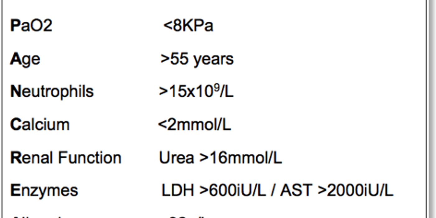
- The criteria are PaO2 below 8 kPa, age over 55, neutrophils above 15 x 10^9/L, calcium below 2 mmol/L, urea above 16 mmol/L, LDH above 600 iU/L or AST above 2000 iU/L, albumin below 32 g/L and glucose above 10 mmol/L.
- Severe acute pancreatitis is defined by persistent organ failure beyond 48 hours.
- Management is supportive: fluids, oxygen, analgesia, and treating the cause.
What Is Acute Pancreatitis?
Acute pancreatitis is a sudden inflammatory condition of the pancreas that can range from a short, mild illness to organ-threatening disease. Acute pancreatitis is sudden inflammation of the pancreas caused by premature activation of pancreatic enzymes, which can injure and autodigest pancreatic tissue. The inflammation may remain localised or trigger a widespread inflammatory response. It can cause serious complications, including shock, respiratory failure, pancreatic necrosis, and organ failure.
The pancreas lies behind the stomach in the upper abdomen. Its exocrine function supports digestion. Acinar cells release enzymes into the pancreatic duct, including amylase, lipase, and proteases. These enzymes digest carbohydrates, fats, and proteins in the small intestine.
Its endocrine function regulates blood glucose. Islet cells release insulin, which lowers blood glucose, and glucagon, which raises it. Severe pancreatic inflammation can temporarily impair both digestive and hormonal functions.
Normally, pancreatic enzymes become active after reaching the intestine. In pancreatitis, enzymes may activate inside the gland. This process causes local tissue injury, oedema, fat necrosis, and inflammation. Gallstones and alcohol are the two commonest causes in the UK.
Pancreatitis inflammation pancreas refers to injury that begins inside the gland when digestive enzymes activate too early. The term pancreatitis inflammation describes the resulting local response, which may extend into nearby tissues or become systemic.
Acute Versus Chronic Disease
An acute episode develops quickly and may settle over days or weeks. The pancreas can recover without permanent structural damage. Chronic pancreatitis causes persistent inflammation, fibrosis, and irreversible loss of exocrine or endocrine function.
Chronic pancreatitis is long-term scarring and functional loss of the gland, whereas acute pancreatitis is a sudden episode that may resolve completely. Repeated acute attacks, alcohol exposure, smoking, and inherited disorders can increase the risk of chronic pancreatitis.
Symptoms of typical symptoms include severe epigastric pain, often radiating to the back, nausea, and vomiting. Patients may prefer sitting forward. Early assessment identifies the cause, confirms the diagnosis, and estimates severity. Initial tests usually include blood tests, serum lipase, liver function tests, calcium, glucose, and imaging when indicated.
This condition is a common and potentially serious gastrointestinal presentation. It accounts for more than 275,000 hospital admissions annually in the United States.
Why It Matters in MRCS Part B
For MRCS Part B, pancreatitis tests applied pathology, physiology, clinical examination, and critical care knowledge. Candidates may need to explain symptoms, interpret tests, recognise systemic inflammation, and begin treatment.
The applied surgical science and critical care station is marked out of 20: 12 marks assess clinical knowledge and application, 4 assess clinical and technical skill, and 4 assess professionalism. Three of the 17 examined stations fall within this area.
Acute pancreatitis is pancreatic inflammation that can progress from local enzyme injury to life-threatening systemic disease, so early assessment and treatment are essential.
Key clinical insight: A patient with suspected acute pancreatitis needs both diagnostic confirmation and an early search for organ dysfunction. A normal early scan does not necessarily exclude clinically important disease.
Exam candidates should connect the diagnosis with practical priorities: recognise the presentation, assess airway and circulation, identify a reversible pancreatitis cause, provide supportive care, and seek senior help when organ failure develops.
What causes acute pancreatitis?
Acute pancreatitis develops when pancreatic enzymes injure the gland before they should become active. Acute pancreatitis is sudden inflammation of the pancreas caused by premature activation of digestive enzymes. Gallstones and alcohol account for most cases in the UK.
Common causes
- Gallstones and alcohol are the two commonest causes of acute pancreatitis in the United Kingdom.
Gallstones can obstruct the common bile duct or pancreatic duct. Ask about biliary colic, previous jaundice, pale stools, dark urine, and post-meal right upper-quadrant pain. Check liver tests and arrange ultrasound tests for gallstones or duct dilatation.
Alcohol-related disease usually follows prolonged, heavy intake. Ask about daily units, binge drinking, dependence, previous withdrawal symptoms, and earlier episodes of pancreatitis. Patients may underreport alcohol use, so ask clearly and without judgement.
- Marked hypertriglyceridaemia, usually above 11.3 mmol/L, can trigger pancreatic inflammation without gallstones or alcohol.
Consider this cause in patients with diabetes, obesity, metabolic syndrome, pregnancy, or a personal history of lipid disorders. Blood tests should include triglycerides when the usual causes are absent.
- Medicines, abdominal trauma, and recent ERCP are recognised causes of acute pancreatic injury.
Review recent prescriptions and dose changes. Reported drug associations include azathioprine, valproate, tetracyclines, and some diuretics. Ask about blunt abdominal trauma, recent surgery, and endoscopic retrograde cholangiopancreatography.
- Infections, anatomical abnormalities, and obstructing lesions can cause pancreatitis when routine investigations are unrevealing.
Consider mumps, viral hepatitis, parasites, pancreas divisum, choledochal cysts, ampullary tumours, and pancreatic malignancy. A family history may suggest hereditary pancreatitis or genetic susceptibility; research into pancreatitis risk factors in children also highlights the importance of inherited and clinical risk factors.
A useful way to remember the differential is to ask whether the cause acute pancreatitis is obstructive, toxic, metabolic, traumatic, infectious, autoimmune, or genetic. This framework also helps when the first ultrasound does not explain the illness.
Common cause acute pancreatitis usually means gallstone disease or alcohol exposure, but a less obvious pancreatitis cause may be important when the episode is recurrent, occurs in a young person, or follows a new medicine.
Clinical risk assessment and MRCS approach
Smoking independently increases pancreatitis risk and may worsen both acute and chronic disease. Autoimmune disease, including systemic lupus erythematosus, and vasculitis are rare causes. Ischaemia from vascular compromise or systemic hypotension can also injure the pancreas.
Use the clinical context to construct a concise differential diagnosis. Start with gallstones and alcohol, then add hypertriglyceridaemia, drugs, trauma, ERCP, infection, and structural disease. Ask targeted history questions before ordering broad tests. This approach supports appropriate treatment and avoids missing a reversible cause.
The phrase causes acute pancreatitis includes both common triggers and unusual mechanisms. For example, a narrowed pancreatic bile duct, a tumour, or a medication reaction may block normal drainage and initiate enzyme injury.
Patients should also be asked about recent drinking, smoking, new medicines, abdominal procedures, family history, and previous attacks. These details can distinguish a first episode from recurrent pancreatitis and can identify people at risk of chronic pancreatitis.
What cause should an MRCS candidate identify first? Identify gallstones or alcohol first, then state the supporting history, examination findings, and targeted tests. If neither fits, name two less common causes and explain your reasoning.
What is the key takeaway? Gallstones and alcohol lead the differential, but history, medication review, triglyceride testing, imaging, and recent procedures reveal less obvious causes.
How does acute pancreatitis present?
Acute pancreatitis commonly presents with persistent upper-abdominal pain, vomiting, and a risk of systemic illness. The problem is sudden inflammation of the pancreas, often causing severe abdominal pain and systemic illness. Presentation ranges from mild, self-limiting disease to pancreatic necrosis, shock, and multiorgan failure. Gallstones and alcohol are the two commonest causes in the UK. Recognising severe symptoms helps guide urgent treatment and escalation.
The typical presentation is severe, constant epigastric or upper abdominal pain. It may spread through to the back and improve when the patient sits forward. Nausea and repeated vomiting are common. Examination may show epigastric tenderness, guarding, reduced bowel sounds, and ileus. Abdominal distension can develop as bowel movement slows.
Pancreatitis acute symptoms can resemble perforated ulcer, cholecystitis, myocardial infarction, bowel obstruction, or mesenteric ischaemia. For that reason, symptom recognition should be followed by an ABCDE assessment rather than relying on pain alone.
Clinical findings and warning signs
Fever and tachycardia suggest inflammation or infection. Hypotension, cold peripheries, oliguria, confusion, or increasing oxygen requirements suggest hypovolaemia or organ dysfunction. Jaundice may indicate a gallstone obstructing the common bile duct. Rarely, Cullen’s sign causes periumbilical bruising, while Grey-Turner’s sign causes flank bruising. These signs may indicate severe retroperitoneal bleeding or necrosis.
A focused MRCS history should establish pain onset, site, radiation, severity, vomiting, fever, jaundice, and bowel function. Ask about alcohol intake, gallstone symptoms, previous episodes, abdominal surgery, trauma, hyperlipidaemia, and medicines. Ask about chest pain and cardiovascular risk factors because myocardial infarction can mimic upper abdominal pain.
Perform an ABCDE assessment before detailed examination. Record observations, hydration, urine output, mental state, and respiratory effort. Examine the abdomen for distension, tenderness, guarding, peritonism, bruising, and bowel sounds. Check the chest, heart, calves, and hernial orifices. This approach demonstrates safe examination technique and identifies patients needing urgent treatment.
Severe acute pancreatitis is defined by persistent organ failure lasting more than 48 hours. Warning features include increasing oxygen requirements, low urine output, kidney dysfunction, hypotension, rising lactate, confusion, and respiratory distress.
A patient with severe acute pancreatitis may need a high-dependency unit or intensive care bed. Early involvement of critical care, gastroenterology, surgery, radiology, and nutrition teams can reduce delays in managing complications.
Severity assessment and differentials
The modified Glasgow-Imrie score is assessed during the first 48 hours. A score of three or more indicates a severe attack. Criteria include:
- PaO₂ below 8 kPa
- Age above 55 years
- Neutrophils above 15 × 10⁹/L
- Calcium below 2 mmol/L
- Urea above 16 mmol/L
- LDH above 600 IU/L or AST above 2000 IU/L
- Albumin below 32 g/L
- Glucose above 10 mmol/L
Symptoms of initial tests include full blood count, urea and electrolytes, liver function tests, calcium, glucose, blood gas, lipase, and inflammatory markers. Ultrasound can identify gallstones and bile duct dilatation. CT assesses complications when the diagnosis is unclear or the patient deteriorates. Early treatment includes oxygen, intravenous fluids, analgesia, antiemetics, monitoring, and nutritional support.
Always consider perforated peptic ulcer, biliary colic or cholecystitis, bowel obstruction, mesenteric ischaemia, ruptured aneurysm, and myocardial infarction. Peritonism, absent bowel sounds, marked distension, or disproportionate pain should prompt urgent reassessment. These alternative diagnoses require different tests and treatment.
Urgent-care insight: Persistent low blood pressure, reduced urine output, confusion, breathlessness, or jaundice should be treated as warning signs rather than expected discomfort from a mild attack.
How Is Acute Pancreatitis Diagnosed?
Diagnosing acute pancreatitis requires two of three findings: characteristic abdominal pain, pancreatic enzymes at least three times the upper limit of normal, or characteristic imaging.
Acute pancreatitis is usually diagnosed when two of three findings are present: typical abdominal pain, pancreatic enzymes at least three times the upper limit of normal, or characteristic imaging. Blood tests also assess organ function, severity, and possible causes.
Confirming the diagnosis
Diagnosis begins with the history and examination. Severe, persistent epigastric pain that may radiate to the back supports the diagnosis. However, symptoms can overlap with perforated ulcer, biliary disease, myocardial infarction, and bowel ischaemia.
The standard definition requires two of three diagnostic findings:
- Typical abdominal pain consistent with pancreatic inflammation.
- Serum lipase or amylase at least three times the upper limit of normal.
- Characteristic findings on ultrasound, CT, or MRI.
Therefore, imaging is not always needed to confirm acute pancreatitis when the pain and blood results are typical. This two-out-of-three approach comes from the revised Atlanta classification.
Serum lipase is generally preferred because it remains raised longer than amylase and is more specific. Amylase may be normal early, late, or in alcohol-related or hypertriglyceridaemic disease. Raised amylase can also occur with renal failure, salivary gland disease, bowel pathology, and some medicines. Enzyme levels do not reliably predict severity.
Initial blood tests should include:
- Full blood count: identifies leucocytosis, haemoconcentration, and possible infection.
- Urea and electrolytes: assess dehydration, renal function, and fluid loss.
- Liver function tests: an early alanine transaminase rise may suggest gallstone disease.
- Calcium: low calcium can indicate more severe inflammation.
- Glucose: detects stress hyperglycaemia or pancreatic endocrine dysfunction.
- Triglycerides: identifies hypertriglyceridaemia as a possible cause.
- C-reactive protein and other inflammatory markers: help monitor the inflammatory response.
- Blood gas and lactate: assess hypoxia, acidosis, and tissue hypoperfusion in unwell patients.
These blood tests support diagnosis and guide early treatment, but no single result confirms or excludes pancreatitis.
Doctors diagnose the condition by combining symptoms, enzyme results, imaging, and clinical severity rather than by relying on one abnormal result. A normal enzyme level does not always exclude disease when the presentation is late or the patient has hypertriglyceridaemia.
Choosing the right imaging test
Imaging should answer a specific clinical question. Transabdominal ultrasound is useful early because it can identify gallstones, bile duct dilatation, and gallbladder disease. Bowel gas and obesity can limit views of the pancreas.
A ct scan is particularly useful when clinicians need to assess complications, uncertain findings, or deterioration. A contrast-enhanced CT scan can show necrosis, collections, haemorrhage, and surrounding tissue injury, although very early imaging may underestimate the extent of disease.
Contrast-enhanced CT provides detailed information about pancreatic inflammation, fluid collections, necrosis, and complications. It is particularly useful when the diagnosis remains uncertain, the patient deteriorates, or complications are suspected. CT may be less informative very early in the illness.
Magnetic resonance cholangiopancreatography (MRCP) gives high-quality images of the bile and pancreatic ducts without endoscopy. It helps detect common bile duct stones when ultrasound is inconclusive. Endoscopic ultrasound can find small stones or sludge and may guide decisions about further treatment. Plain chest or abdominal radiographs are not diagnostic, but can identify alternative diagnoses or complications.
The term endoscope refers to a flexible instrument used to view or access internal structures. During ERCP, an endoscope reaches the duodenum so clinicians can treat obstruction in the bile ducts or pancreatic bile duct when indicated.
Assessing severity and cause
Severity assessment starts immediately. Clinicians monitor blood pressure, heart rate, oxygenation, urine output, renal function, and evidence of persistent organ failure. Severe pancreatitis involves organ failure lasting more than 48 hours and may require critical care support.
The modified Glasgow-Imrie score is calculated during the first 48 hours. A score of three or more indicates a severe attack. The criteria are:
- PaO₂ below 8 kPa
- Age over 55 years
- Neutrophils above 15 × 10⁹/L
- Calcium below 2 mmol/L
- Urea above 16 mmol/L
- LDH above 600 IU/L or AST above 2000 IU/L
- Albumin below 32 g/L
- Glucose above 10 mmol/L
Symptoms of other tools include the Ranson score, APACHE II, and the BISAP score. These tools support risk assessment but never replace repeated clinical review. Diagnosis must also identify the cause, especially gallstones, alcohol use, medicines, high triglycerides, and metabolic disease.
The safest diagnostic approach combines two-of-three confirmation, targeted imaging, cause-focused blood tests, and early assessment for organ failure.
How is acute pancreatitis managed?
Acute pancreatitis is treated initially with supportive hospital care, close monitoring, appropriate nutrition, and management of the underlying trigger.
What is the initial treatment?
Acute pancreatitis treatment is mainly supportive during the first hours. Start with an ABCDE assessment, two intravenous lines, regular observations, urine output, and repeated blood tests. Check full blood count, urea and electrolytes, glucose, calcium, liver function, C-reactive protein, blood gas, and triglycerides.
Give oxygen if hypoxic, aiming for an oxygen saturation of 94–98% in most adults. Use a lower target of 88–92% for patients at risk of carbon dioxide retention. Treat severe pain with titrated intravenous opioids, while considering paracetamol where suitable. Antiemetics can control vomiting and improve comfort.
Give balanced intravenous crystalloid for hypovolaemia. Reassess pulse, blood pressure, capillary refill, urine output, lung signs, and haematocrit frequently. Avoid indiscriminate fluid loading, particularly in older patients or those with cardiac or renal disease. A urinary catheter may help measure output in severe pancreatitis.
Acute pancreatitis treated promptly with fluids, symptom control, nutritional support, and monitoring may resolve without an invasive procedure. However, severe acute pancreatitis requires repeated reassessment because organ dysfunction can evolve after the initial presentation.
Why are monitoring and nutrition important?
The inflammatory response can cause shock, kidney injury, respiratory failure, and abdominal compartment syndrome. Escalate to a high-dependency or intensive care setting if there is persistent hypotension, hypoxia, oliguria, rising lactate, confusion, or organ failure.
Severity assessment uses clinical findings, organ function, imaging, and serial tests. The modified Glasgow-Imrie score is calculated during the first 48 hours. A score of 3 or more indicates a severe attack. Its criteria include:
- PaO₂ below 8 kPa
- Age above 55 years
- Neutrophils above 15 × 10⁹/L
- Calcium below 2 mmol/L
- Urea above 16 mmol/L
- LDH above 600 IU/L or AST above 2000 IU/L
- Albumin below 32 g/L
- Glucose above 10 mmol/L
Start oral feeding when nausea, vomiting, and pain improve. Do not keep patients nil by mouth without a clear reason. If oral intake is not possible, provide early enteral feeding, usually through a nasogastric tube. Post-pyloric feeding may help when gastric feeding is poorly tolerated. Parenteral nutrition is reserved for situations where enteral feeding is impossible.
Treatment considerations include avoiding unnecessary fasting and avoiding excessive fluid administration. Nutrition, renal function, respiratory status, and tolerance of oral intake should be reviewed throughout admission.
What is the role of antibiotics and procedures?
Routine prophylactic antibiotics do not prevent sterile pancreatic necrosis and can promote resistance, fungal infection, and adverse effects. Use antibiotics for proven or strongly suspected infection, such as cholangitis, infected necrosis, pneumonia, urinary infection, or sepsis from another source. Infected necrosis may present with fever, deterioration, gas within a collection, or positive culture.
Urgent ERCP is indicated for biliary obstruction with cholangitis, and for persistent obstruction causing ongoing jaundice. It is not routine treatment for every gallstone-related attack. In mild gallstone pancreatitis, perform laparoscopic cholecystectomy during the same admission, ideally before discharge.
A narrowed pancreatic bile duct may require specialist evaluation with MRCP, endoscopic ultrasound, or ERCP. During a suitable procedure, an endoscope can help remove an obstructing stone or place a stent to restore drainage.
Delay cholecystectomy after severe disease if significant collections or necrosis remain. In frail patients who cannot tolerate surgery, ERCP with sphincterotomy may provide an alternative, but risks require specialist review.
When is surgery or drainage needed?
Most necrosis and fluid collections need observation, not immediate operation. Drain or debride infected necrosis using a step-up approach: antibiotics, image-guided or endoscopic drainage, then minimally invasive necrosectomy if required. Open surgery is reserved for selected cases.
The procedure may involve endoscopic drainage, image-guided drainage, or minimally invasive removal of dead tissue. Clinicians generally avoid early open surgery because operating before collections mature can increase complications.
Urgent multidisciplinary care is needed for abdominal compartment syndrome, uncontrolled haemorrhage, bowel ischaemia or perforation, pancreatic fistula, persistent sepsis, or symptomatic mature pseudocysts. The team may include surgeons, gastroenterologists, intensivists, interventional radiologists, microbiologists, and nutrition specialists.
The key treatment principle is early supportive care, selective ERCP, timely gallbladder removal, and delayed minimally invasive intervention for complications.
This quick-reference table summarises key acute pancreatitis treatment priorities during the initial hours, including assessment, monitoring, oxygen targets, symptom control, intravenous fluids, and laboratory evaluation based on the guidance outlined in this section.
Treating acute pancreatitis therefore means more than controlling symptoms. Clinicians must identify whether gallstones, a narrowed pancreatic duct, alcohol, metabolic disease, or another trigger requires a specific intervention.
If a gallstone blocks the pancreatic bile duct and causes cholangitis, urgent ERCP may be required. An endoscope can access the bile ducts, remove the obstruction, and sometimes place a stent. Later gallbladder removal can help prevent another episode.
What are the complications of acute pancreatitis?
Acute pancreatitis can produce local collections, necrosis, organ failure, nutritional problems, and recurrent disease. Acute pancreatitis is inflammation of the pancreas that can cause local tissue damage, systemic inflammation, and temporary or permanent organ failure. Mild pancreatitis usually settles within one week. Severe pancreatitis needs close monitoring and may require intensive care.
Complications and recovery
Symptoms of local complications include pancreatic necrosis, fluid collections, pseudocysts, and walled-off necrosis. A pseudocyst is a mature, fluid-filled collection. Walled-off necrosis contains fluid and dead pancreatic tissue, usually developing after several weeks.
Dead tissue can become infected and may require drainage or delayed necrosectomy. Severe cases are managed by a multidisciplinary team that balances infection control, nutrition, organ support, and the timing of any procedure.
Necrosis can become infected, causing fever, worsening pain, sepsis, or organ deterioration. Treatment often involves antibiotics for suspected infection, drainage, and minimally invasive necrosectomy. A delayed, step-up approach is generally preferred when the patient is stable.
Venous thrombosis may affect the splenic, portal, or superior mesenteric veins. Bleeding can result from vessel erosion or a pseudoaneurysm. A sudden haemodynamic decline, gastrointestinal bleeding, or falling haemoglobin requires urgent CT angiography and specialist intervention. Embolisation is often the definitive treatment.
Symptoms of systemic complications include persistent organ failure, respiratory failure, renal failure, shock, and multiorgan dysfunction. Increased intra-abdominal pressure can cause abdominal compartment syndrome, with reduced urine output, hypotension, and ventilatory difficulty. This requires urgent senior review, bladder-pressure measurement, and sometimes surgical decompression.
Most patients with mild pancreatitis recover fully. Recovery from severe pancreatitis may take weeks or months. Fatigue, altered bowel function, and reduced appetite can continue after discharge. Follow-up should assess nutrition, pain, glucose control, and recurrent symptoms.
After discharge from hospital, patients may need review of weight, stool changes, glucose, alcohol intake, smoking, and medication risks. Persistent diarrhoea, weight loss, oily stools, or new diabetes can suggest exocrine or endocrine dysfunction and should prompt clinical review.
Acute and chronic disease
Acute pancreatitis causes sudden inflammation and severe, persistent epigastric pain. Chronic pancreatitis causes long-term fibrosis, with recurrent or continuous pain that may eventually lessen as the pancreas loses function.
Chronic disease can cause malabsorption, weight loss, steatorrhoea, and diabetes. The pancreas may no longer produce enough digestive enzymes or insulin. Long-term treatment can include pancreatic enzyme replacement, nutritional support, diabetes care, analgesia, and selected endoscopic or surgical procedures.
Chronic pancreatitis treated successfully requires a long-term plan rather than care for one isolated episode. Management may include enzyme replacement, dietary support, diabetes monitoring, smoking cessation, alcohol cessation, and specialist procedures for duct obstruction or persistent symptoms.
Repeated episodes of acute pancreatitis can increase the likelihood of chronic pancreatitis, especially when the trigger continues. Chronic pancreatitis can also predispose to nutritional deficiencies and impaired quality of life.
Prevention and MRCS revision checklist
Prevention depends on the cause. Patients should stop or reduce alcohol, receive definitive gallstone treatment when appropriate, and review possible medication triggers. Manage severe hypertriglyceridaemia, avoid smoking, and arrange follow-up after recurrent attacks. Repeated inflammation increases the risk of chronic pancreatitis.
Preventing acute pancreatitis means addressing the trigger: remove or treat gallstones when clinically appropriate, avoid harmful drinking, stop smoking, manage triglycerides, and review medicines with a clinician.
Patients should not attempt to remove prescribed medicines independently. A clinician should assess whether a drug is a plausible trigger and decide whether an alternative is safer. Preventing recurrent episodes can also help prevent chronic pancreatitis.
For MRCS revision, link each complication to the following response:
- Shock, confusion, low urine output: assess ABCDE, repeat blood tests, arterial blood gas, lactate, electrolytes, and renal function; escalate to critical care.
- Respiratory distress: check oxygen saturation, chest imaging, and blood gas; provide oxygen or ventilation support.
- Necrosis or collections: request contrast CT when clinically appropriate; involve a specialist pancreatic team; drain or debride infected or symptomatic collections.
- Bleeding: identify haemodynamic instability and falling haemoglobin; obtain CT angiography; arrange urgent embolisation or surgery.
- Gallstone pancreatitis with cholangitis or persistent obstruction: arrange urgent ERCP, followed by cholecystectomy when safe.
- Recurrent attacks: investigate alcohol, gallstones, medications, triglycerides, calcium, and structural disease.
Can one attack cause chronic pancreatitis? Usually not, but severe or recurrent pancreatitis can damage the gland and lead to permanent exocrine or endocrine failure.
Recognising organ failure, necrosis, bleeding, and infection early improves escalation, treatment, recovery, and prevention of chronic pancreatic disease.
How is this topic marked in MRCS Part B?
MRCS Part B marks an applied surgical science and critical care station out of 20: 12 marks for clinical knowledge and its application, 4 for clinical and technical skill and 4 for professionalism. Three of the seventeen examined stations fall in this area.