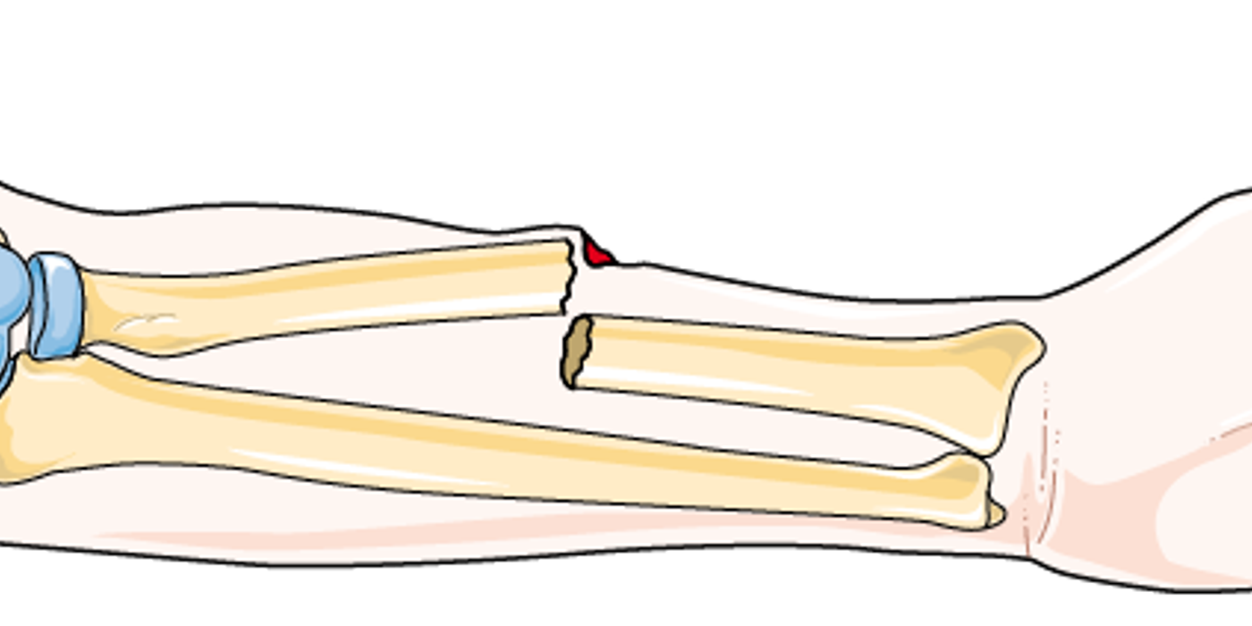
Open fractures require emergency care because a broken bone communicates with the outside environment, creating a high risk of infection and further damage. Protect the open wound, assess circulation, apply a sterile dressing and splint, give urgent antibiotics and tetanus protection, and arrange specialist surgical treatment.
An open fracture is a time-critical emergency: do not push exposed bone back inside, probe the wound, or delay transfer for detailed classification.
Key takeaways
- The Gustilo-Anderson classification runs I, II, IIIA, IIIB and IIIC, and is assigned properly at debridement rather than from the wound on arrival.
- Type I is a wound under 1 cm with minimal soft tissue damage; type II is over 1 cm with moderate damage; type III is high-energy with extensive damage, and IIIC carries an arterial injury.
- BOAST: antibiotics within one hour of injury; debridement immediate for gross contamination or vascular compromise, within 12 hours for high-energy, within 24 hours for low-energy.
- Definitive soft tissue cover within 72 hours if it is not achieved at debridement.
- Photograph the wound, remove gross contamination only, cover with a saline-soaked dressing, splint the limb, and give tetanus prophylaxis.
What does defining open fractures involve?
Defining open fractures means identifying communication between a broken bone and the external environment, even when the skin opening is small or distant from the break. A compound fracture and an open fracture describe the same clinical problem. Dirt, clothing fibres, water, or faecal material can enter through an open wound and increase the risk infection.
The Gustilo-Anderson fracture classification is a wound-severity classification confirmed during operative assessment. Clinicians still use the Gustilo-Anderson fracture type to guide antibiotics, fixation, debridement, and soft-tissue cover.
What is an open fracture?
An open fracture is a broken bone connected to the outside environment through a wound in the skin or soft tissues. It is also called a compound fracture. The wound may be directly over the break or several centimetres away. Even a small puncture can expose the fracture to bacteria.
This injury can damage muscles, tendons, nerves, blood vessels, and surrounding skin. It is therefore both a fracture and a soft-tissue emergency.
- Any fracture with a wound communicating with the outside environment should be treated as an open injury until specialist assessment excludes it.
- The skin wound may occur away from the fracture, so its location does not reliably show the underlying bone injury.
- Contamination from soil, clothing, saliva, or farm environments increases infection risk and influences urgent antibiotic and surgical management.
- The Gustilo-Anderson system classifies these injuries as types I, II, IIIA, IIIB, or IIIC after surgical debridement.
- Type III injuries involve high-energy trauma, extensive soft-tissue damage, contamination, vascular injury, or the need for flap coverage.
A compound fracture is not the same as a closed fracture. A closed fracture has no communication with the exterior, whereas compound fractures may expose bone, fracture haematoma, or deeper structures to bacteria. The injury zone can include the skin, muscle, nerves, vessels, and an open joint.
A tibia fracture is a common example because the tibia has limited soft-tissue coverage along part of its length. However, any bones can be involved. A compound injury can also affect the femur, humerus, forearm, hand, foot, pelvis, or ankle.
Can a small wound still indicate a serious injury? Yes. The wound size does not always reflect the depth of contamination, tissue damage, or vascular compromise. Never probe, wash deeply, or push exposed bone back into the wound.
Treat every suspected open fracture as a contaminated limb-threatening injury requiring urgent assessment, protection, antibiotics, and specialist care.
Why does prompt management change the outcome?
An open fracture has no direct health benefits. The broken bone communicates with the outside environment, allowing contamination and tissue damage. The injury may also involve blood vessels, nerves, muscles, or joints. Infection risk rises with wound severity.
The benefits come from rapid, structured treatment. Early antibiotics, wound protection, splinting, surgical debridement, and specialist care can reduce preventable harm. This approach helps protect soft tissue, improve control of contamination, support bone healing, and preserve limb function. It also allows earlier movement and rehabilitation when the wound and fracture are stable.
Benefits of prompt management
Effective treatment provides several measurable advantages:
- Lower infection risk: Antibiotics and thorough debridement reduce bacterial contamination before it becomes deep infection.
- Better soft-tissue survival: Saline-soaked dressings and splinting limit further injury to skin, muscle, and exposed bone.
- Improved fracture healing: Stable fixation supports alignment and may shorten the time to union.
- Better functional recovery: Early, safe joint movement reduces stiffness and supports rehabilitation.
- Shorter hospital stays: Definitive internal fixation has been linked with reduced hospital time compared with definitive external fixation.
- Faster return of function: Internal fixation has also been associated with shorter time to union and improved functional outcomes.
These benefits depend on injury severity and treatment timing. A type I wound differs greatly from a type IIIB or IIIC injury. Severe wounds may need flap coverage or vascular repair. The classification is confirmed during surgical assessment, not from wound size alone.
How does the Gustilo-Anderson fracture type affect treatment?
The Gustilo-Anderson fracture type helps clinicians estimate infection risk, soft-tissue loss, and reconstructive requirements. A Gustilo-Anderson fracture type III includes high-energy damage, extensive contamination, or vascular compromise. Within this group, a Gustilo-Anderson fracture type iii may require coordinated orthopaedic, vascular, and plastic surgery.
A fracture type is not determined reliably by a photograph alone. The surgeon assesses the injury zone, wound contamination, devitalised structures, and bone coverage during debridement. This distinction prevents underestimating complex wounds.
Do open fractures heal better than closed fractures? No. They usually carry greater infection and soft-tissue risks. Prompt antibiotics, tetanus prophylaxis, debridement, fixation, and coverage improve the chance of healing.
For an MRCS Part B station, state the practical priorities: photograph the wound, remove gross contamination, cover it with saline-soaked gauze, assess circulation and sensation, splint the limb, and give antibiotics and tetanus prophylaxis.
Rapid, coordinated treatment does not benefit the fracture itself; it reduces infection, protects tissue, and improves healing and function.
How do you manage an open fracture at first presentation?
Treat open fractures as limb-threatening injuries until proven otherwise. Follow trauma priorities, assess circulation and sensation, cover and splint the wound, then arrange urgent antibiotics and operative management.
What is the initial treatment?
The initial treatment for an open fracture is trauma assessment, bleeding control, wound protection, neurovascular examination, splinting, antibiotics, tetanus protection, and urgent specialist referral. Emergency care should address life-threatening problems before detailed limb examination.
Start with immediate priorities
- Activate the trauma pathway and follow ATLS principles. Most patients need emergency department assessment because other serious injuries may coexist.
- Record the mechanism, time, location and environment of injury. Ask whether the wound involved soil, water, farm material, a high-energy impact or a gunshot. These details help estimate contamination and guide antibiotic treatment.
- Assess the limb’s neurovascular status before and after every intervention. Document pulses, capillary refill, skin colour, temperature, motor function and sensation. Also check for worsening pain or pain on passive stretch, which may suggest compartment syndrome.
- Photograph the wound if local policy permits, before applying a dressing. Avoid repeated examination or unnecessary handling. Wound size and severity are classified more accurately during formal surgical debridement.
- Remove only obvious gross contamination that can be lifted away safely. Do not probe the wound or push exposed bone back inside. A formal operation is required for thorough irrigation and debridement.
What does injury stabilisation involve?
Injury stabilisation involves covering the open wound, supporting the limb, and maintaining circulation while definitive care is arranged. Splinting reduces movement at the fracture site and can limit additional damage to nerves, vessels, and soft tissue.
Protect the limb and prevent infection
- Cover the injury with sterile gauze soaked in saline. This reduces drying and limits further contamination while the patient awaits definitive care. Do not seal a heavily contaminated wound without specialist advice.
- Splint the limb above and below the suspected fracture. Immobilisation reduces pain, bleeding and further soft-tissue damage. Recheck circulation and sensation after applying the splint.
- Administer broad-spectrum intravenous antibiotics as soon as possible, according to local guidance. Give tetanus prophylaxis when indicated. Immediate antibiotics and urgent irrigation with debridement form the foundation of early treatment.
- Arrange urgent orthopaedic review for operative debridement and fracture stabilisation. Severe wounds may require early transfer to a specialist centre. Plastic surgery input is often needed for durable soft-tissue coverage.
- Classify the injury using the Gustilo-Anderson system at debridement. Type I is a clean wound under 1 cm. Type II measures 1–10 cm without extensive soft-tissue damage. Type IIIA involves extensive injury but allows bone coverage. Type IIIB needs flap coverage. Type IIIC includes vascular injury requiring repair.
The Gustilo-Anderson fracture type is provisional before surgery and may change after full exploration. For example, a suspected type II may become fracture type iii when deep muscle loss, severe wound contamination, or vascular damage is identified. The Gustilo-Anderson fracture type III includes IIIA, IIIB, and IIIC patterns.
Never use wound size alone to dismiss a compound fracture. The injury zone and depth of contamination may be more important than the visible opening. The first response is simple: assess circulation, cover the wound, splint the limb, give antibiotics and tetanus protection, and obtain urgent specialist care.
What is current best practice?
Best practice combines early antibiotics, careful wound protection, formal debridement, stable fixation, and coordinated soft-tissue reconstruction. The correct plan depends on the patient’s physiology, fracture pattern, injury zone, and wound contamination.
What should happen first?
An open fracture is a fracture that communicates with the outside environment through a wound. Treat any fracture with a nearby wound as open until proven otherwise. The wound may not lie directly over the broken bone.
Use an ATLS approach before focusing on the limb. Assess breathing, circulation, bleeding, and other injuries.
For the affected limb:
- Record pulses, capillary refill, sensation, and motor function.
- Check for ongoing bleeding and compartment syndrome.
- Photograph the wound before dressing it, where local policy allows.
- Remove only obvious gross contamination.
- Cover it with sterile, saline-soaked gauze.
- Apply a splint above and below the suspected fracture.
- Give intravenous antibiotics and assess tetanus protection.
Repeat and document neurovascular findings after splinting. A threatened or ischaemic limb requires immediate specialist and vascular input.
Why do classification and timing matter?
The Gustilo-Anderson grade is assigned after surgical assessment and debridement. It helps predict contamination, soft-tissue damage, and reconstructive needs. Infection risk rises with severity. These figures vary with injury pattern and treatment quality.
Research from the American Academy of Orthopaedic Surgeons and evidence reviews available support early antimicrobial treatment, careful classification, and multidisciplinary reconstruction. The Tscherne system and work by Harald Tscherne may also help describe soft-tissue damage, although the Gustilo-Anderson system remains central for open fractures.
How should definitive treatment be organised?
Definitive treatment requires operative irrigation, debridement, fracture stabilisation, and durable soft-tissue cover. Debridement removes devitalized tissue and foreign material that could support infection or nonunion.
Start antibiotics as soon as possible, following local antimicrobial guidance. Arrange urgent transfer to an orthopaedic trauma centre when specialist care is unavailable. Current evidence supports urgent, planned debridement rather than automatic emergency surgery for every case. Severe contamination, vascular compromise, or an agricultural injury needs faster intervention.
Definitive care combines skilled debridement, stable fracture fixation, and early soft-tissue coverage. Orthopaedic and plastic surgery teams should plan these stages together.
What happens during debridement?
Debridement is the surgical removal of contaminated, devitalized, or non-viable material from the open fracture. The team examines the entire injury zone, identifies viable muscle and bone, removes foreign material, and irrigates under controlled conditions.
An intraoperative wound assessment may reveal deeper damage than initial imaging suggests. For severe compound fractures, surgeons may plan a second look, repeat debridement, external fixation, or staged reconstruction. The goal is to preserve viable structures while reducing bacterial load.
Primary amputation is uncommon but may be considered when the limb is nonviable or reconstruction cannot provide a safe, functional result. Amputation decisions require senior orthopaedic, vascular, plastic surgery, and rehabilitation input.
What is the safest first dressing? Sterile gauze soaked with saline, applied after removing gross contamination. What is the key MRCS answer? “Assess and document neurovascular status, give antibiotics and tetanus prophylaxis, cover and splint the limb, then arrange specialist debridement and fixation.”
Photograph, cover, splint, medicate, document neurovascular status, and arrange urgent specialist treatment.
What are the common mistakes?
Open fractures are broken bones that communicate with the outside environment through a wound, even when the wound is not directly over the break. Treat any fracture with an associated wound as open until proven otherwise.
Common first-aid mistakes
Do not push exposed bone back under the skin. Avoid probing, repeatedly opening, or scrubbing the wound. These actions can introduce further contamination and damage soft tissue.
Do not wash the wound with soap or high-pressure fluid outside a controlled surgical setting. Evidence suggests that adding soap increases the risk of reoperation. Remove only obvious surface contamination, then cover the injury with sterile, saline-soaked gauze.
Do not leave the limb unsupported. Splint above and below the suspected fracture. Check and document pulses, capillary refill, sensation, and movement before and after splinting.
Do not delay emergency transfer while trying to obtain perfect photographs or a detailed classification. Photograph the wound once, if local policy allows, then cover it. The Gustilo-Anderson grade is assigned after surgical assessment and debridement, not from the initial appearance alone.
Treatment errors to avoid
Do not forget early intravenous broad-spectrum antibiotics or tetanus assessment. These measures reduce infection risk and should begin as soon as possible. Also assess for vascular injury and compartment syndrome.
Do not assume a small wound means a minor injury. The wound may lie away from the fracture, and high-energy injuries can cause extensive internal damage. Severe injuries may require orthopaedic, vascular, and plastic surgery input.
Do not postpone specialist debridement unnecessarily.
Do not confuse external fixation with definitive care in every case. External fixation may provide temporary alignment for unstable compound fractures, while later fixation and soft-tissue cover support healing. Imaging helps define the fracture pattern, but it does not replace operative assessment.
Complications include deep infection, osteomyelitis, nonunion, malunion, compartment syndrome, vascular compromise, stiffness, chronic pain, and amputation. A tibia fracture has particular risk because limited coverage can complicate healing.
The safest approach is to cover, splint, assess circulation, give antibiotics and tetanus prophylaxis, and arrange urgent specialist care without manipulating the wound.
What are the BOAST timings for an open fracture?
In the UK the timings come from the British Orthopaedic Association Standard for Trauma (BOAST) for open fractures. Intravenous prophylactic antibiotics should be given as soon as possible, ideally within one hour of injury. Debridement is immediate for highly contaminated wounds — agricultural, aquatic or sewage — and for those with vascular compromise; within 12 hours for other solitary high-energy open fractures; and within 24 hours for all other low-energy open fractures. Definitive soft tissue closure or coverage should be achieved within 72 hours of injury if it is not done at the time of debridement. Quoting 'within six hours' is the commonest way to lose the mark here: that standard was superseded.
How is this topic marked in MRCS Part B?
MRCS Part B marks the procedural stations out of 20. Procedural skills with a patient carries 8 marks for clinical and technical skill and 4 each for clinical knowledge, communication and professionalism. Procedural skills (technical), performed on a model, carries 12 marks for skill and 8 for knowledge, with no communication or professionalism marks. Two of the seventeen examined stations are procedural.