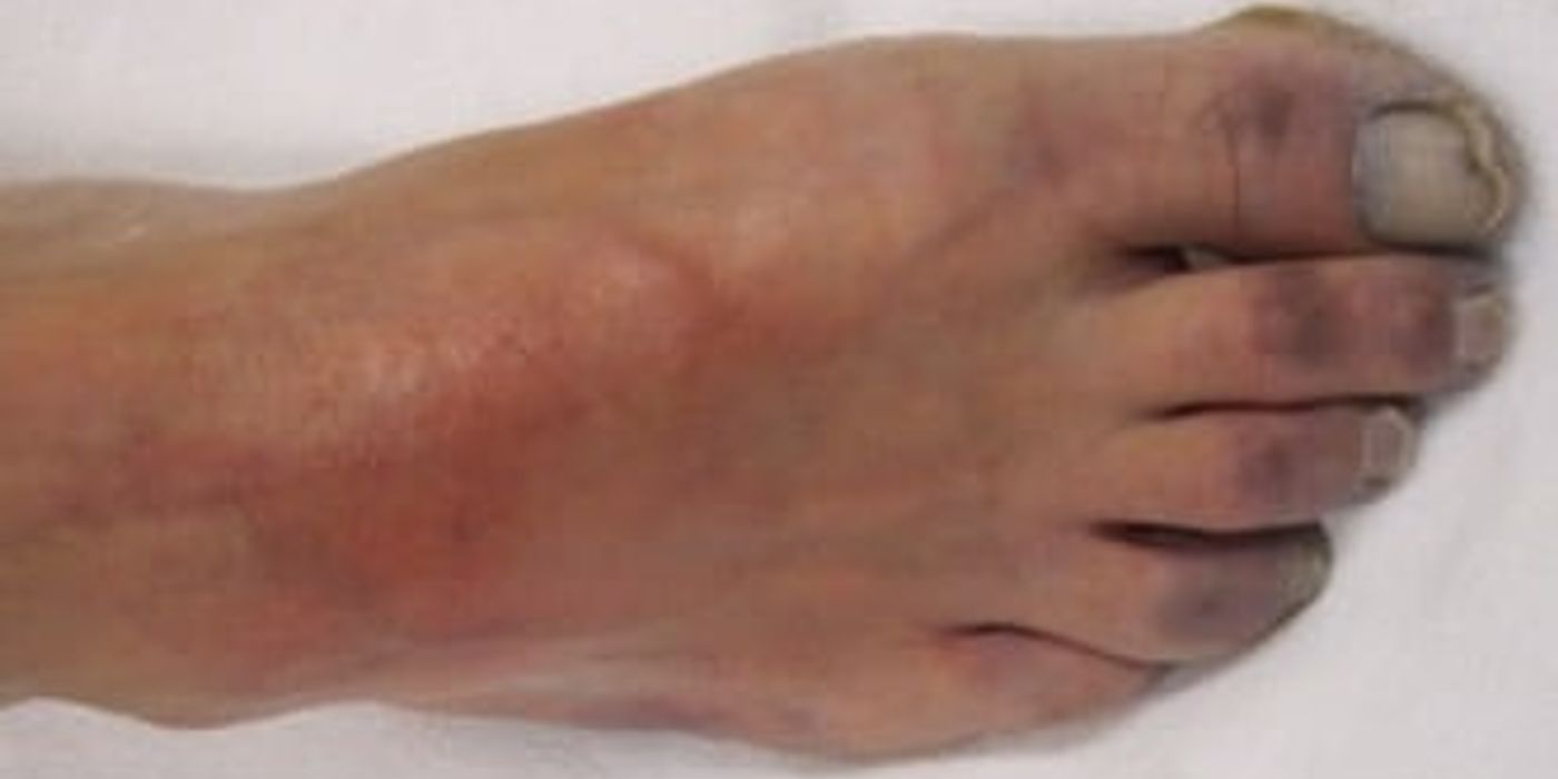
Acute limb ischaemia is a time-critical loss of blood flow that can permanently damage nerves and muscle within hours. Immediate clinical assessment, intravenous heparin when appropriate, and urgent revascularization by thromboembolectomy, endovascular therapy, or surgery can preserve function and life.
Key takeaways
- The six Ps: pain, pallor, pulselessness, perishing cold, paraesthesia and paralysis.
- Paraesthesia and paralysis mark a threatened limb that needs immediate revascularisation.
- Embolus and in-situ thrombosis are the two mechanisms, and the history separates them: sudden onset with a source against a background of claudication.
- Give heparin and analgesia while you arrange imaging and definitive treatment.
- Irreversible muscle damage follows prolonged ischaemia, so time to revascularisation is the thing that matters.
What is acute limb ischaemia?
Acute limb ischaemia is a sudden reduction in arterial blood flow that threatens the viability of a limb. The fall in perfusion causes tissue ischaemia and may lead to irreversible muscle and nerve injury. This condition usually develops within 14 days of symptom onset. It requires urgent assessment and treatment.
ALI is an emergency syndrome caused by inadequate perfusion of a lower extremity or upper extremity. The immediate diagnostic question is whether the tissues are viable, threatened, or already irreversibly injured. This severity classification guides anticoagulation, imaging, revascularization, and the need for emergency care.
Chronic limb ischaemia develops gradually, usually from peripheral arterial disease. Collateral vessels may partly maintain perfusion despite arterial narrowing. Patients often report exertional symptoms, such as intermittent claudication, rather than sudden pain.
Acute-on-chronic ischaemia occurs when an already compromised limb loses further blood flow. A thrombosed atherosclerotic artery, graft or stent can cause this deterioration. Existing collateral circulation may make the symptoms less dramatic, but the limb remains at risk.
The severity of arterial ischaemia depends on the artery involved, the completeness of occlusion and the duration of reduced flow. Occlusion of a major artery produces more extensive ischaemia than blockage of a smaller branch. A sudden, complete obstruction allows little time for collateral circulation to compensate.
Irreversible ischaemic injury can develop within hours, particularly when the limb has poor collateral supply. Nerves and skeletal muscle are especially vulnerable. Paraesthesia and paralysis suggest a threatened limb and require immediate revascularisation. Fixed mottling, profound anaesthesia or rigor may indicate irreversible ischaemia.
Clinical insight: A normal-looking foot does not exclude ALI. Collateral arteries, early presentation, and partial obstruction can preserve colour while nerve function is deteriorating.
Definition and clinical context
Chronic limb ischaemia develops gradually, usually from peripheral arterial disease. Collateral vessels may partly maintain perfusion despite arterial narrowing. Patients often report exertional symptoms, such as intermittent claudication, rather than sudden pain.
Acute-on-chronic ischaemia occurs when an already compromised limb loses further blood flow. A thrombosed atherosclerotic artery, graft or stent can cause this deterioration. Existing collateral circulation may make the symptoms less dramatic, but the limb remains at risk.
The severity of arterial ischaemia depends on the artery involved, the completeness of occlusion and the duration of reduced flow. Occlusion of a major artery produces more extensive ischaemia than blockage of a smaller branch. A sudden, complete obstruction allows little time for collateral circulation to compensate.
Irreversible ischaemic injury can develop within hours, particularly when the limb has poor collateral supply. Nerves and skeletal muscle are especially vulnerable. Paraesthesia and paralysis suggest a threatened limb and require immediate revascularisation. Fixed mottling, profound anaesthesia or rigor may indicate irreversible ischaemia.
Causes of acute arterial occlusion
The main mechanisms are embolism and in-situ thrombosis:
- Embolism: A clot travels from the heart, commonly with atrial fibrillation, recent myocardial infarction or prosthetic valve disease. It may lodge at an arterial bifurcation.
- In-situ thrombosis: Thrombosis forms within an artery affected by atherosclerosis. A history of claudication or disease in the opposite limb supports this cause.
- Graft or stent occlusion: Previous vascular intervention can become thrombosed or stenosed.
- Trauma: Arterial laceration, compression or intimal injury can reduce distal perfusion.
- Arterial dissection or aneurysm: A dissecting flap, thrombosed aneurysm or distal embolisation can obstruct flow.
Symptoms of typical symptoms include sudden pain, pallor, pulselessness, a perishing cold limb, paraesthesia and paralysis: the six Ps. The presentation varies with the affected artery and the speed of occlusion.
ALI is a vascular and surgical emergency because untreated ischaemia progresses to tissue necrosis, limb loss, systemic complications and death. Acute limb ischaemia is a time-critical arterial emergency in which rapid recognition and revascularisation protect limb and life.
Which anatomy matters in acute limb ischaemia?
The occlusion level predicts examination findings and influences the revascularization strategy. A clot in the common femoral artery can remove flow to the entire lower extremity, whereas disease in the popliteal artery may preserve femoral pulses but eliminate pedal signals.
A thrombosed popliteal aneurysm can create both local obstruction and distal emboli. A popliteal aneurysm may therefore present with foot ischaemia even when the original aneurysm is not immediately obvious on examination.
When imaging shows disease throughout the femoropopliteal artery, the team may need thromboembolectomy, bypass, catheter therapy, or a hybrid approach. An occlusion involving the entire femoropopliteal artery usually requires a more detailed plan than an isolated embolus.
How Does Acute Limb Ischaemia Present?
Acute limb ischaemia is a sudden reduction in arterial blood flow that threatens tissue survival. It requires rapid assessment and treatment.
Recognising the six Ps
- Pain is usually sudden and severe, reflecting inadequate oxygen delivery to muscles and nerves during acute limb ischaemia.
- Pallor occurs when reduced arterial flow leaves the limb pale, although dependent positioning may produce dusky discoloration.
- Pulselessness suggests arterial obstruction, but chronic ischaemia may also cause absent pulses through established peripheral arterial disease.
- Perishing cold describes a cool or cold limb, especially when compared with the opposite side during examination.
- Paraesthesia indicates sensory nerve dysfunction and suggests worsening ischaemia requiring urgent vascular assessment and possible revascularisation.
- Paralysis is a late, ominous sign of advanced ischaemia and indicates a threatened limb needing immediate emergency treatment.
Pain alone does not define severity. Progressive sensory loss, weakness, or paralysis indicates worsening nerve and muscle ischaemia. Fixed mottling, muscle rigidity, and profound neurological loss suggest irreversible injury. Muscle damage may become irreversible after approximately 6 hours, although timing varies with collateral circulation and the underlying cause.
Practical examination approach
- Compare both limbs for colour, temperature, capillary refill, sensation, movement, and pain before interpreting an isolated vascular finding.
- Palpate femoral, popliteal, posterior tibial, and dorsalis pedis pulses, documenting the last palpable pulse and comparing both sides.
- Assess capillary refill and skin temperature, remembering that delayed refill and a cold limb support poor distal perfusion.
- Use handheld Doppler to assess arterial and venous signals; absent arterial signals with neurological deficit indicates severe ischaemia.
- Test light touch, pinprick, and motor function at the ankle and toes, because sensory loss precedes paralysis in many cases.
- Ask about symptom onset, atrial fibrillation, myocardial infarction, claudication, malignancy, trauma, and previous arterial disease.
An embolus often causes abrupt, complete ischaemia in a previously well limb. Atrial fibrillation, recent myocardial infarction, or a prosthetic valve may provide the source. In-situ thrombosis often develops in a diseased artery, with previous claudication and possible symptoms in the opposite limb.
A chronically ischaemic limb may remain warm and pink because collateral arteries have developed. Therefore, a pulse deficit alone does not confirm an acute emergency.
What clinical features suggest a threatened extremity?
The most important clinical features are new sensory loss, motor weakness, absent Doppler signals, and progressive skin change. These findings have greater urgency than symptom intensity alone. A patient with modest discomfort but new foot drop may need faster intervention than a patient with intense symptoms and preserved neurology.
Emergency care pathways commonly combine bedside examination, Rutherford severity classification, point-of-care Doppler, and rapid consultation with a vascular surgeon. The diagnostic process should remain focused: determine viability, identify the likely obstruction, and select therapy without avoidable delay.
A distal embolus may be treated with thromboembolectomy when the occlusion is accessible. If an angiogram shows total occlusion in a diseased segment, the team may choose thrombus aspiration, CDT, angioplasty, or bypass rather than isolated catheter extraction.
When is immediate senior vascular input required? Call urgently for any acute limb ischaemia with paraesthesia, weakness, paralysis, severe pain, marked pallor, fixed mottling, or absent Doppler signals. Give analgesia, obtain intravenous access, administer oxygen when clinically indicated, and start anticoagulation according to local protocol while arranging urgent revascularisation.
MRCS examination tip: describe findings systematically, escalate early, and never delay vascular review for unnecessary investigations in a threatened limb. A cold, painful limb with sensory or motor loss is limb-threatening ischaemia until proven otherwise.
How do you assess the severity of the ischaemia?
The challenge in acute limb ischaemia is identifying whether the limb remains viable. Delayed recognition can allow reversible ischaemia to progress to nerve and muscle death. Assess pain, colour, temperature, capillary refill, sensation, movement, and pulses. The classic six Ps are pain, pallor, pulselessness, perishing cold, paraesthesia, and paralysis.
Severity classification is the bedside process of deciding whether tissue is viable, threatened, or irreversible. Rutherford classification remains the central severity classification for triage, although clinical judgement and serial examinations are also essential.
The direct answer is to use the Rutherford classification: Paraesthesia and paralysis indicate threatened ischaemia. Paralysis, including new foot drop, signals severe nerve ischaemia and requires immediate action. Fixed mottling suggests irreversible ischaemia.
Bedside assessment and investigations
Start with bilateral pulse palpation and handheld arterial Doppler. An absent arterial signal supports the diagnosis and helps localise the occlusion. An absent venous signal, usually at the popliteal vein, indicates advanced ischaemia and a poor prognosis. If only a Doppler signal is audible, a perfusion pressure below 50 mmHg suggests severe ischaemia.
An ankle-brachial pressure index (ABPI) can quantify arterial perfusion when the patient is stable and measurements are possible. However, pain, calcified vessels, and severe ischaemia can make ABPI unreliable. Bedside Doppler findings and the clinical examination take priority. Never delay treatment for ABPI, formal ultrasound, or other tests when the limb is immediately threatened.
CT angiography (CTA) is usually the preferred imaging test for locating the artery blockage. It maps inflow, collateral circulation, and distal run-off. This helps the vascular team choose embolectomy, thrombolysis, angioplasty, stenting, or bypass. CTA is appropriate when it will not delay revascularisation. The same principles apply to acute limb ischaemia (spelled ischemia in US sources), .
Send blood for full blood count, urea and electrolytes, glucose, coagulation studies, group and save, and creatine kinase. Check lactate and blood gas results when severe ischaemia or systemic illness is suspected. These tests assess tissue injury and prepare for intervention, but they do not confirm or exclude limb ischaemia.
If an embolus is suspected, perform an ECG for atrial fibrillation or recent myocardial infarction. Arrange cardiac examination and echocardiography to identify atrial thrombus, ventricular thrombus, valve disease, or another embolic source. This assessment must occur alongside urgent limb treatment, not instead of it.
Rutherford classification determines urgency: diagnose the limb at the bedside, image selectively, and never let investigations delay revascularisation for threatened ischaemia.
What does imaging add to the diagnostic process?
Imaging identifies the level, length, and mechanism of obstruction when it can be obtained without delaying treatment. Duplex ultrasound can assess flow and aneurysm, while CTA provides a rapid anatomical map for surgical and endovascular planning.
An angiogram may be performed during intervention. If the angiogram shows total occlusion, the operator can evaluate distal run-off and decide whether thromboembolectomy, percutaneous thrombus aspiration, CDT, or surgical revascularization is most appropriate.
The differential diagnosis includes venous thrombosis, compartment syndrome, vasospasm, aortic dissection, and trauma. A focused diagnostic pathway helps clinicians avoid unnecessary testing while preserving time for emergency care.
How is acute limb ischaemia managed?
Acute limb ischaemia is a vascular emergency. Give analgesia, intravenous unfractionated heparin, fluids when needed, and obtain urgent vascular review while arranging rapid revascularisation for a threatened limb.
Immediate treatment
Acute limb ischaemia means sudden arterial obstruction that reduces blood flow to viable tissue. Treat suspected ischaemia as an emergency, even before imaging confirms the artery involved. The clinical priorities are to preserve the limb, control pain, prevent clot extension, and identify the cause.
Call the vascular team immediately and involve anaesthesia, interventional radiology, and critical care when required. Record the onset of symptoms and examine both limbs. Document pulses, capillary refill, skin colour, temperature, sensation, and motor function. Reassess frequently because ischaemia can progress quickly.
Give intravenous unfractionated heparin unless there is a clear contraindication, such as active major bleeding or heparin-induced thrombocytopenia. Heparin prevents further thrombus propagation but does not remove the existing obstruction. Follow local protocols for the initial bolus and infusion, with monitoring of clotting results.
Provide adequate analgesia and establish intravenous access. Give intravenous fluids if the patient is dehydrated or hypotensive. Administer oxygen if hypoxic, rather than routinely to every patient. Keep the limb warm, but avoid direct heat because an ischaemic limb can burn easily. Position the limb dependent or flat; avoid elevation, which may further reduce arterial perfusion.
The classic six Ps are pain, pallor, pulselessness, perishing cold, paraesthesia, and paralysis. Paraesthesia and paralysis suggest a threatened limb and require immediate revascularisation. Fixed mottling, profound anaesthesia, and rigor indicate advanced tissue injury and possible irreversible ischaemia.
Choosing revascularisation
The treatment depends on the cause, the severity of ischaemia, the artery involved, and the patient’s operative risk. Revascularisation may be surgical, endovascular, or hybrid.
An embolectomy removes an embolus, often using a balloon catheter through an exposed artery. It is particularly suitable for a sudden occlusion without severe underlying arterial disease. Thrombectomy removes clot from a locally diseased artery, but additional angioplasty, stenting, or bypass may be necessary.
Thromboembolectomy is the removal of an embolus or thrombus using an open catheter-based technique. A Fogarty catheter is passed beyond the obstruction, inflated, and withdrawn to restore flow. Thromboembolectomy is often effective for an embolus at the common femoral or popliteal artery, but it may not correct the underlying artery disease.
Thromboembolectomy can be followed by completion angiography to identify residual clot, dissection, stenosis, or distal embolisation. A second thromboembolectomy may be required when clot fragments remain. The decision should account for the Rutherford category and the patient’s operative risk.
In-situ thrombosis often reflects advanced peripheral arterial disease. Endovascular treatment can restore flow with catheter thrombectomy, aspiration, angioplasty, stenting, or mechanical clot disruption. Bypass may provide more durable flow when disease is extensive or the artery is unsuitable for catheter treatment. Surgical and endovascular methods can also be combined.
Percutaneous thrombus aspiration uses negative pressure through a catheter to remove clot without an open incision. It may be useful for selected occlusions in the femoropopliteal segment, particularly when rapid flow restoration is needed.
Percutaneous thrombus aspiration and thrombus aspiration can be used as part of a staged endovascular procedure. The operator may combine aspiration with balloon angioplasty, stenting, or CDT when residual stenosis remains after clot removal.
Catheter-directed thrombolysis delivers a fibrinolytic drug directly into the clot through a catheter. It may be considered for a viable or marginally threatened limb, particularly when thrombosis is distal, extensive, or difficult to treat surgically. It is generally unsuitable for a suddenly paralysed limb because treatment is too slow. Major active bleeding, recent intracranial haemorrhage, recent major surgery, uncontrolled hypertension, and significant bleeding risk are important contraindications.
CDT, or catheter-directed thrombolysis, delivers clot-dissolving medication directly into an occluded vessel. CDT may be selected when the obstruction is extensive and the patient is stable enough for monitored therapy.
A second CDT strategy may follow incomplete aspiration or thromboembolectomy. However, CDT should never delay emergency revascularization in Rutherford IIb disease, where neurological loss indicates threatened tissue.
A threatened limb requires rapid restoration of arterial flow, not delayed investigation or isolated treatment. The final plan should be coordinated by vascular surgery, interventional radiology, anaesthesia, and critical care teams.
The key management principle is simple: anticoagulate immediately, involve vascular specialists urgently, and revascularise before a threatened limb becomes irreversibly ischaemic.
Which treatment is used for popliteal aneurysm thrombosis?
A thrombosed popliteal aneurysm requires treatment of both the clot and the aneurysm. Isolated thromboembolectomy may restore temporary flow but leave a continuing source of embolisation or thrombosis.
For thrombosed popliteal aneurysm, options include bypass exclusion, thromboembolectomy, CDT, percutaneous thrombus aspiration, or a hybrid repair. The popliteal artery and distal run-off should be assessed carefully because emboli may obstruct tibial arteries.
A thrombosed popliteal lesion can be managed with surgical revascularization when anatomy is complex or tissue is immediately threatened. A second surgical revascularization procedure may be necessary if initial flow restoration does not provide adequate distal perfusion.
When do you revascularise, and what goes wrong afterwards?
What complications follow revascularisation?
Reperfusion injury is tissue damage caused when blood flow returns to an ischaemic limb. Oxygen free radicals, oedema and inflammatory mediators increase capillary leakage. This can worsen swelling after correcting acute limb ischaemia.
The major local complication is compartment syndrome. Muscle swelling raises pressure inside a closed fascial compartment. Arterial inflow then falls again, causing recurrent ischaemia despite palpable pulses.
Suspect compartment syndrome with:
- Increasing pain, especially on passive stretch
- Tense, swollen compartments
- New paraesthesia or weakness
- Pain poorly controlled by analgesia
Pulses may remain present, so a pulse does not exclude this complication. In severe ischaemia lasting more than six hours, assess carefully for compartment syndrome. Some guidance supports prophylactic fasciotomy, although evidence and practice vary.
Why are metabolic abnormalities dangerous?
Reperfusion releases potassium, lactate, myoglobin and acidic metabolites into the circulation. This may cause hyperkalaemia, metabolic acidosis, rhabdomyolysis and acute kidney injury.
Check potassium, bicarbonate, creatine kinase, blood gas and renal function soon after reperfusion. Hyperkalaemia can cause life-threatening cardiac arrhythmias. Treat severe abnormalities urgently using local emergency protocols.
Maintain intravenous fluids while monitoring urine output. Dark urine suggests myoglobinuria. A falling urine output or rising creatinine may indicate renal injury. Severe hyperkalaemia or renal failure may require renal replacement therapy.
How is the surgical plan chosen?
Revascularisation may involve embolectomy, thrombectomy, thrombolysis, angioplasty, bypass or a hybrid procedure. The choice depends on the artery, cause, duration of ischaemia, limb viability and patient fitness.
Surgical revascularization restores blood flow through an open operation such as bypass or thromboembolectomy. It is often preferred for immediately threatened tissue, accessible emboli, failed catheter therapy, or anatomy unsuitable for an endovascular approach.
A hybrid repair combines open surgical revascularization with endovascular therapy. For example, a surgeon may perform femoral thromboembolectomy before balloon angioplasty of a stenosed femoropopliteal segment.
Assess fasciotomy needs before and after restoring flow. Decompress all clinically threatened compartments. Leave wounds open when swelling is substantial, with delayed closure or skin grafting later.
Amputation is appropriate when the limb is irreversibly ischaemic, non-functional or cannot be safely revascularised. Fixed mottling, rigor, profound anaesthesia and paralysis suggest irreversible ischaemia. Do not revascularise clearly dead tissue, because reperfusion can trigger severe systemic toxicity. Discuss palliation when surgery offers no meaningful benefit.
What requires postoperative monitoring?
Perform regular documented checks of:
- Limb colour, temperature and capillary refill
- Pulses, Doppler signals and graft or artery patency
- Pain, sensation, movement and compartment tension
- Potassium, acid–base status, creatine kinase and urine output
- Incisions, fasciotomy wounds, bleeding and infection
Recurrent pain, paraesthesia, paralysis or loss of a Doppler signal suggests renewed ischaemia. Wound bleeding may indicate anticoagulation-related haemorrhage or technical failure.
How is recurrence prevented?
Continue anticoagulation when embolism, atrial fibrillation or another thrombotic indication is present. Antiplatelet therapy is commonly used after arterial intervention or for atherosclerotic disease. Control smoking, diabetes, hypertension and lipid levels.
Investigate the embolic source with electrocardiography, cardiac monitoring and echocardiography when appropriate. Consider atrial fibrillation, recent myocardial infarction, valve disease, aneurysm and intracardiac thrombus.
Long term management reduces recurrent obstruction and protects the opposite extremity. It may include statin therapy, antiplatelet treatment, anticoagulation for atrial fibrillation, supervised exercise, diabetes control, smoking cessation, and review of artery disease.
Follow-up may combine duplex surveillance, medication review, walking assessment, and specialist vascular review. The exact therapy depends on whether the original event was embolic, thrombotic, aneurysm-related, or associated with chronic peripheral artery disease.
Revascularisation treats the artery, but careful monitoring prevents reperfusion injury, compartment syndrome, renal failure and avoidable limb loss.
What else causes a cold, painful limb?
Acute limb ischaemia is a sudden reduction in arterial blood flow that threatens limb viability. It usually causes severe pain, pallor, pulselessness, a perishing cold limb, paraesthesia and paralysis. Paraesthesia or paralysis suggests advanced ischaemia and requires immediate vascular involvement.
Distinguishing the main differentials
Deep vein thrombosis usually causes a warm, swollen and tender limb. Pulses remain present, although oedema may make them difficult to feel. Arterial ischaemia instead produces a cold, pale limb with reduced or absent pulses. Severe venous obstruction can cause phlegmasia.
Compartment syndrome causes severe, escalating pain and pain on passive muscle stretch. The limb may feel tense, but distal pulses can remain present. Paraesthesia may develop from nerve compression. In arterial ischaemia, pulselessness, pallor and coldness are more prominent.
Acute-on-chronic peripheral arterial disease often has a slower onset. The patient may describe previous claudication, rest pain, ulcers or reduced walking distance. The opposite limb may also have weak pulses and signs of vascular disease. Chronic collateral vessels can make the affected limb less dramatically pale.
Venous phlegmasia causes massive swelling, cyanosis and pain from extensive venous thrombosis. The limb may become threatened by arterial inflow compromise. Look for marked oedema rather than an isolated cold, pulseless limb. This is a vascular emergency requiring urgent specialist assessment.
Embolus, thrombosis and unusual causes
An embolus often causes sudden, severe symptoms in a previously normal limb. There may be no history of claudication, and the opposite limb may have normal pulses. Ask about atrial fibrillation, recent myocardial infarction, valve disease or a cardiac murmur. Cardiac disease is a common embolic source.
In-situ thrombosis usually develops on a background of atherosclerosis. Symptoms may progress over hours, with previous claudication and abnormal pulses in the contralateral limb. Both mechanisms cause ischaemia, so the history supports—but does not replace—vascular imaging.
Trauma raises concern for arterial transection, intimal disruption or thrombosis. Iatrogenic injury may follow catheterisation, arterial puncture or orthopaedic procedures. Obtain urgent vascular advice and consider CT angiography if the patient is stable. Do not delay revascularisation in a threatened limb for unnecessary imaging.
Aortic dissection can obstruct an arterial branch and produce unequal pulses or blood pressures. Chest, back or abdominal pain increases suspicion. This changes treatment: involve vascular and cardiothoracic teams urgently, because treating the limb alone may miss a lethal aortic cause.
MRCS prioritisation framework
- Identify immediate threats: assess airway, breathing and circulation, while recognising limb-threatening ischaemia.
- Examine both limbs: compare colour, temperature, sensation, power and pulses from femoral to pedal vessels.
- Find the mechanism: assess cardiac risk factors, atherosclerosis, trauma and recent procedures.
- Act promptly: give analgesia, intravenous fluids where appropriate, and systemic heparin unless contraindicated. Seek urgent vascular review.
In a cold, painful limb, absent pulses favour arterial ischaemia, but the six Ps, history and bilateral examination distinguish the cause and determine urgency.
What is acute limb ischaemia, and why is it a vascular emergency?
Acute limb ischaemia is a sudden reduction in arterial blood flow that threatens tissue survival. It develops when an artery becomes blocked by an embolus, thrombosis, trauma, or another vascular problem. Without oxygen, nerves and muscles develop ischaemia, causing pain, pallor, coldness, and neurological symptoms. The limb can become irreversible within hours, especially when collateral circulation is poor. This is a medical emergency requiring urgent vascular assessment and treatment. Give analgesia, intravenous fluids, oxygen when indicated, and intravenous unfractionated heparin unless contraindicated.
What are the six Ps of acute limb ischaemia?
The six Ps are pain, pallor, pulselessness, perishing cold, paraesthesia, and paralysis. Pain is often severe and sudden. Pallor and a cold limb indicate reduced perfusion, while absent pulses suggest arterial occlusion. Paraesthesia shows sensory nerve dysfunction and signals a threatened limb. Paralysis is a late, serious sign of motor nerve and muscle ischaemia. Examine both limbs, compare pulses, assess capillary refill, and use a handheld Doppler. Do not rely on the six Ps alone, because early ischaemia may have subtle symptoms. Progressive neurological loss requires immediate senior and vascular review.
How is limb viability assessed using the Rutherford classification?
The Rutherford classification grades the limb from viable to irreversibly damaged according to sensory loss, muscle weakness, and Doppler signals. Category I is viable, with no sensory or motor deficit and audible arterial and venous signals. Category IIa is marginally threatened, with limited sensory loss but no weakness. Category IIb is immediately threatened, with sensory loss beyond the toes and motor weakness; urgent revascularisation is required. Category III is irreversible, with profound anaesthesia, paralysis, absent arterial and venous signals, and established tissue damage. Fixed mottling supports irreversible ischaemia and carries a poor prognosis.
When are heparin, embolectomy, or thrombolysis used?
Heparin is given immediately, while embolectomy or thrombolysis depends on the cause, severity, and duration of ischaemia. Intravenous heparin prevents clot extension while treatment is arranged. An embolus causing a sudden occlusion in a previously healthy artery often requires urgent surgical embolectomy, particularly in Rutherford IIb ischaemia. Catheter-directed thrombolysis may suit selected patients with a viable or marginally threatened limb, especially when thrombosis affects diseased distal vessels. It is slower and unsuitable for an immediately threatened limb or major bleeding risk. Angioplasty or bypass may treat underlying arterial disease.
What is the difference between an embolus and arterial thrombosis?
An embolus travels from another site, whereas arterial thrombosis forms directly within a diseased artery. Emboli commonly arise from atrial fibrillation, recent myocardial infarction, or prosthetic valves. They cause abrupt, severe ischaemia without previous claudication, and the opposite limb may have normal pulses. In-situ thrombosis usually develops on atherosclerotic plaque or an aneurysm. The patient may describe preceding claudication, a slower deterioration, and disease in the contralateral limb. Ask about arrhythmia, cardiac symptoms, vascular history, malignancy, dehydration, and prothrombotic conditions.
What complications can occur after revascularisation?
Reperfusion can cause compartment syndrome, metabolic disturbance, bleeding, renal injury, and recurrent ischaemia. Swollen muscle compartments can raise pressure and impair microcirculation, causing worsening pain, pain on passive stretch, sensory loss, or weakness. Urgent four-compartment fasciotomy may be required. Reperfusion can also release potassium, acid, and myoglobin, producing hyperkalaemia, acidosis, arrhythmia, and acute kidney injury. Heparin and thrombolysis increase bleeding risk, including gastrointestinal or retroperitoneal haemorrhage. Monitor pulses, limb temperature, neurological function, urine output, electrolytes, and creatine kinase after treatment.
How should MRCS Part B candidates practise this topic?
Candidates should practise a structured vascular examination and explain management using a clear emergency sequence. Start with the six Ps, compare both limbs, assess pulses from femoral to pedal level, and use Doppler signals. State the Rutherford category, begin immediate resuscitation, prescribe heparin, and request urgent vascular help. Then explain the likely embolic or thrombotic cause and definitive treatment. The applied surgical science and critical care area contains three of the 17 examined stations: 12 marks assess clinical knowledge, four assess clinical and technical skill, and four assess professionalism. Mrcspartbquestions provides over 3,500 interactive questions, examination scenarios, explanations, and mark schemes for structured practice.
For MRCS Part B, recognise threatened ischaemia early, classify the limb accurately, and communicate decisive management.
How is this topic marked in MRCS Part B?
MRCS Part B marks an applied surgical science and critical care station out of 20: 12 marks for clinical knowledge and its application, 4 for clinical and technical skill and 4 for professionalism. Three of the seventeen examined stations fall in this area.