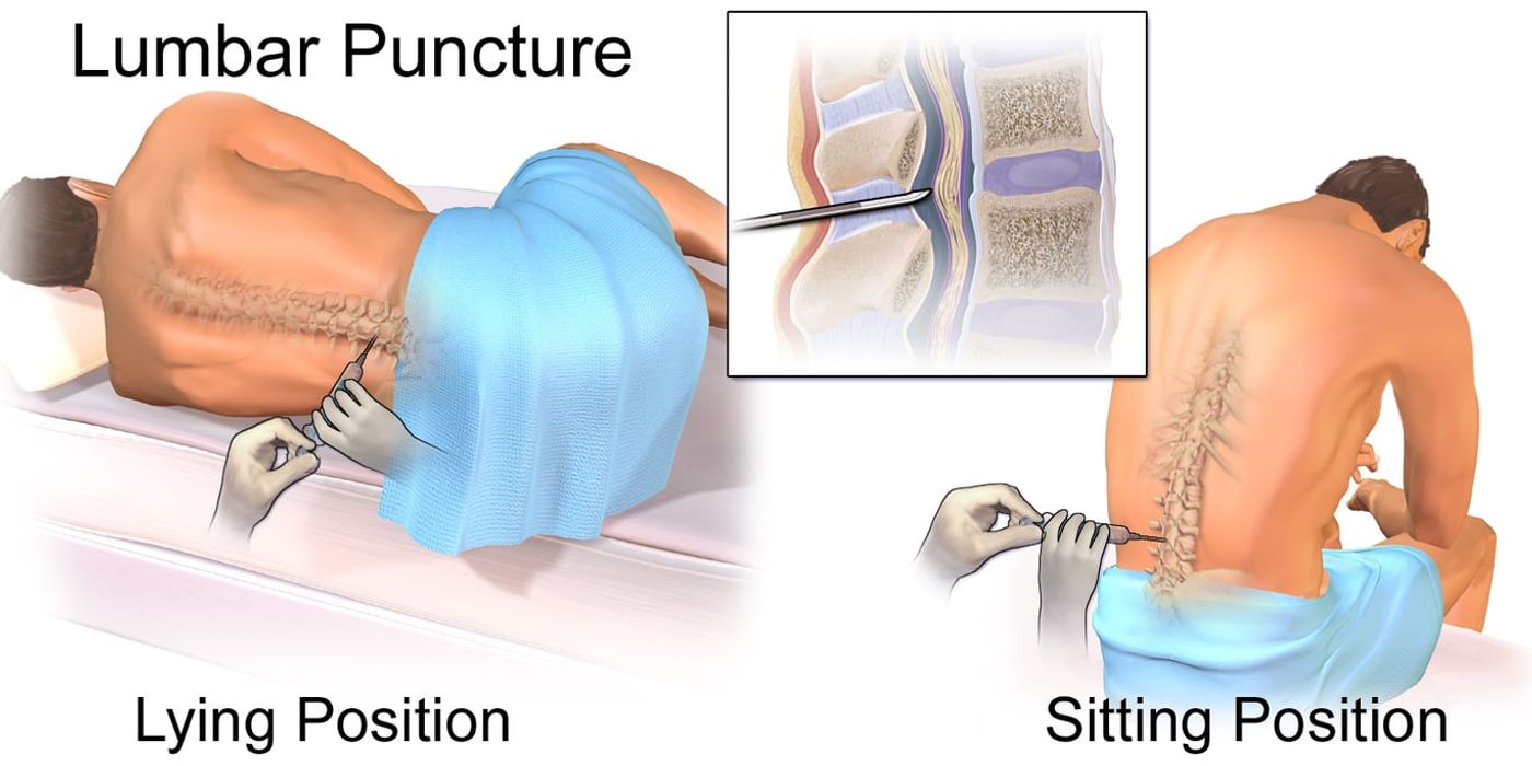
A lumbar puncture, also called a spinal tap, collects a small sample of cerebrospinal fluid from the lower back for laboratory testing. The test remains an important way to investigate meningitis, bleeding, inflammation, cancer, and selected neurological conditions; local anaesthetic usually makes it tolerable, although pressure, brief discomfort, or a later headache can occur.
A lumbar puncture is performed below the usual end of the spinal cord, so the needle normally does not enter the cord itself.
Key takeaways
- Position the patient in the left lateral fetal position with the back at the edge of the bed, or sitting and leaning forward.
- Insert at the L3/L4 or L4/L5 interspace, below the termination of the spinal cord at L1/L2.
- The needle passes through skin, subcutaneous fat, supraspinous ligament, interspinous ligament, ligamentum flavum, dura and arachnoid.
- Measure the opening pressure before collecting samples, and send them in numbered bottles.
- The opening pressure usually taught is 10 to 20 cmH2O; a prospective adult series gives a wider clinical range of 6 to 25 cmH2O.
What is a lumbar puncture, and why is it done?
A lumbar puncture is a diagnostic procedure that removes cerebrospinal fluid (CSF) from the lower spinal canal for testing. Lumbar puncture is a diagnostic procedure that removes cerebrospinal fluid (CSF) from the lower spinal canal for testing.
CSF, also called spinal fluid, is a clear fluid surrounding the brain and spinal cord. It cushions the central nervous system and helps maintain its chemical environment. A hollow spinal needle collects a small fluid sample from the subarachnoid space.
The lumbar region provides safer access because the spinal cord usually ends at the L1–L2 level. The needle enters below this point, usually through the L3/L4 or L4/L5 interspace. In the left lateral fetal position, the patient’s back should be at the edge of the bed.
From superficial to deep, the needle passes through the skin, subcutaneous fat, supraspinous ligament, interspinous ligament, ligamentum flavum, dura, and arachnoid. The needle then reaches the spinal subarachnoid space, where CSF flows.
Lumbar puncture spinal tap is the common patient-facing description of this test. Spinal tap lumbar puncture means the same diagnostic examination, while lumbar puncture LP is a shortened clinical abbreviation. These terms describe sampling rather than spinal anaesthesia or an epidural.
Why Is It Performed?
A diagnostic spinal procedure may help investigate:
- Suspected meningitis or other central nervous system infection
- Inflammatory disease, including multiple sclerosis
- Subarachnoid haemorrhage, when initial imaging is inconclusive
- Selected neurological conditions affecting the spinal or brain systems
- Abnormal pressure or cellular, chemical, and microbiological changes in spinal fluid
The fluid is sent for analysis. Tests may include opening pressure, cell count, protein, glucose, Gram stain, culture, and further specialised studies.: What It Is, Purpose & Procedure)
A diagnostic lumbar puncture differs from spinal anaesthesia. Spinal anaesthesia deliberately injects an anaesthetic into spinal fluid to cause temporary numbness. A therapeutic spinal procedure may remove fluid or deliver treatment. These purposes must not be confused during consent or examination.
For MRCS Part B, candidates should link applied spinal anatomy with indications, contraindications, and communication. Discuss infection, bleeding risk, anticoagulants, abnormal coagulation, and possible raised intracranial pressure. Explain benefits, discomfort, bleeding, infection, headache, and rare neurological complications clearly.
Does the needle enter the spinal cord? Usually, no. The needle enters below the spinal cord’s usual termination, reducing this risk. A lumbar puncture safely samples spinal fluid below the spinal cord to diagnose infection, inflammation, bleeding, and selected neurological disease.
How do you assess, consent and prepare the patient?
Before a spinal tap, the clinician confirms the indication, reviews safety risks, and explains how the test will be performed. Before a lumbar puncture, the clinician confirms the indication, reviews safety risks, and explains the spinal procedure clearly. The aim may be to obtain cerebrospinal fluid (CSF), measure pressure, or deliver spinal medication.
Clinical assessment and safety checks
- A complete spinal assessment reviews symptoms, neurological findings, medications, allergies, infection, bleeding risk, and relevant imaging before the procedure.
- Raised intracranial pressure from a mass lesion or obstructed fluid pathway can make spinal needle insertion dangerous.
- Coagulopathy, anticoagulant treatment, thrombocytopenia, or local infection may contraindicate the procedure until corrected or treated.
The clinician asks about headache, fever, seizures, altered consciousness, weakness, sensory change, and symptoms of raised intracranial pressure. They perform a focused neurological examination, including conscious level, pupils, limb power, sensation, reflexes, and gait when appropriate.
The the medication history must include anticoagulants and antiplatelet drugs. Check allergies, especially to local anaesthetic, antiseptic agents, dressings, and latex. Review platelet count and coagulation results. Normal clotting must be confirmed before spinal needle insertion, where clinically indicated.
Look for cellulitis or another infection at the proposed puncture site. Consider brain imaging when there are focal neurological signs, papilloedema, reduced consciousness, new seizures, or concern about a mass lesion. The purpose is to reduce the risk of brain herniation, bleeding, or infected CSF.
Consent and patient preparation
Explain why CSF fluid is needed and how the procedure will occur. Discuss expected pressure or discomfort from local anaesthetic and needle passage. Explain possible post-procedure headache, back discomfort, bleeding, infection, traumatic fluid sampling, and failure to obtain fluid. Rare complications include nerve irritation, persistent spinal headache, or brain herniation in unsuitable patients.
Obtain informed consent before the spinal procedure. Confirm identity, indication, allergies, relevant results, and equipment. Prepare an assistant, monitoring equipment, sterile gloves, antiseptic solution, local anaesthetic, dressings, and suitable spinal needles.
Position the patient in the left lateral fetal position, with the back at the edge of the bed. Flex the lumbar spine without obstructing breathing. Identify the L3/L4 or L4/L5 interspace, below the spinal cord’s usual termination at L1/L2.
The needle passes through skin, subcutaneous fat, supraspinous ligament, interspinous ligament, ligamentum flavum, dura, and arachnoid before entering CSF fluid.
MRCS Part B focus
In the MRCS Part B surgical skills station, present this as a structured spinal procedure: assess, consent, position, identify landmarks, maintain sterility, and explain complications. Procedural stations are marked out of 20 marks. A patient-based station awards 8 for clinical and technical skill, plus 4 each for knowledge, communication, and professionalism.
#### FAQ Why must raised intracranial pressure be considered? A spinal procedure can create a pressure gradient and precipitate fatal brain herniation when an obstructing lesion is present.
What is the key preparation principle? Do not puncture until the indication, neurological safety, coagulation, infection risk, consent, and spinal anatomy have been checked.
Safe spinal fluid sampling begins with assessment, informed consent, correct positioning, and a clearly explained procedure. Lumbar puncture spinal safety depends on checking coagulation, reviewing medicines, and assessing symptoms that could indicate raised intracranial pressure. A puncture spinal tap should be delayed when additional tests or specialist review are required.
Clinicians may use ultrasound or fluoroscopy-assisted planning for difficult anatomy, although the basic safety checks remain essential. Additional tests can include a platelet count, coagulation studies, blood glucose, neuroimaging, or microbiology requested before the sample is collected.
How do you perform a lumbar puncture?
A lumbar puncture is performed with the patient positioned safely, the skin cleaned, local anaesthetic administered, and a sterile needle advanced into the lower subarachnoid space.
A lumbar puncture can seem concerning because a needle enters the lower spinal region. Patients often worry about spinal injury, pain, fluid leakage, or headache. The procedure is performed below the end of the spinal cord, reducing the risk of direct cord injury. A trained clinician must follow local policy, use suitable supervision, and confirm that the procedure is appropriate.
The procedure removes cerebrospinal fluid (CSF) from the spinal canal using sterile technique and local anaesthetic. The patient usually lies in the left lateral fetal position, with the back at the edge of the bed. The seated position may be used when necessary. In either position, spinal flexion opens the lumbar spaces.
Positioning, preparation, and needle insertion
- The clinician introduces themselves, confirms consent, checks the indication, and prepares an assistant. The patient’s observations and relevant safety checks should be completed before the spinal procedure.
- In the lateral position, the patient curls forwards while keeping the shoulders and hips aligned. In the seated position, the patient leans forwards over a support. Excessive twisting can make the spinal landmarks harder to identify.
- A line between the highest points of the posterior iliac crests approximates the L4 level. The insertion site is usually the L3/L4 or L4/L5 interspace. These sites are below the spinal cord’s usual termination at L1/L2.
- The clinician cleans the skin using an appropriate antiseptic solution and applies sterile drapes. Aseptic technique is essential because infection can follow contamination. Circular application of povidone-iodine is one recommended approach.
- Local anaesthetic is injected into the skin and deeper tissues. The clinician waits for its effect before advancing the spinal needle. Patients may feel pressure, but sharp pain should be reported immediately.
The spinal needle passes through the skin, subcutaneous fat, supraspinous ligament, interspinous ligament, ligamentum flavum, dura, and arachnoid. It then enters the subarachnoid space, where spinal fluid surrounds the nerves. A trained clinician uses anatomical landmarks to guide needle placement.
Fluid collection and aftercare
If clinically required, opening pressure is measured with the patient relaxed in the lateral position. CSF is then allowed to drip into several numbered containers. Each container is labelled at the bedside and sent promptly for the requested fluid tests, such as cell count, glucose, protein, microbiology, or other investigations.
The clinician replaces the stylet before withdrawing the spinal needle. A small sterile dressing covers the puncture site. The patient is observed according to local policy, including for worsening headache, back pain, bleeding, infection, or neurological symptoms. Persistent headache or new spinal symptoms require clinical review.
Who performs the procedure? A suitably trained clinician performs this spinal procedure under appropriate supervision. The exact equipment, spinal needle, fluid handling, pressure measurement, and aftercare must follow local policy.
A safe lumbar puncture uses correct spinal positioning, strict asepsis, a low lumbar entry point, careful fluid collection, and supervised clinical technique.
Lumbar puncture spinal technique uses a low entry point because the spinal cord usually ends above the selected interspace. During a puncture spinal tap, the clinician confirms that the needle inserted below the cord is in the intended space.
The needle inserted for a lumbar puncture may be a cutting or atraumatic design, depending on local policy. A stylet is replaced before withdrawal to reduce the chance of a post-procedure leak. If the sample does not flow, the clinician should not repeatedly advance or redirect without reassessing position and safety.
What Does a Lumbar Puncture Feel Like?
A lumbar puncture usually feels like a brief sting followed by pressure or pushing, rather than severe pain, because an anaesthetic numbs the skin and deeper tissues.
Local anaesthetic usually makes the procedure tolerable, but you may feel a brief sting, pressure, pushing, or tingling. Staying still and reporting sharp pain helps the healthcare provider complete the spinal tap safely.
During the procedure
A lumbar puncture usually causes less pain than patients expect. The local anaesthetic first produces a brief sting. It then numbs the skin and deeper tissues before the spinal needle enters.
You may feel pressure, pushing, or mild tingling as the needle advances. These sensations do not necessarily mean something is wrong. Tell the healthcare provider immediately if you feel sharp, burning, or electric pain.
The procedure is usually performed with you lying in the left lateral fetal position. Your back stays close to the edge of the bed. Curling forward widens the spaces between the vertebrae.
The needle passes into the lower spinal canal, commonly through the L3/L4 or L4/L5 interspace. This site lies below the usual end of the spinal cord at L1/L2. The spinal needle passes through several layers before reaching the cerebrospinal fluid.
Cerebrospinal fluid (CSF) is the clear fluid surrounding the brain and spinal cord. It flows through the needle into collection tubes for laboratory testing. Remaining still helps the healthcare provider control the needle and collect fluid safely. Sudden movement can make the procedure harder or require repositioning. Slow breathing and clear communication can help you stay relaxed.
You may feel pressure while the needle enters, although many people feel little or no pain.
Immediately afterwards
Mild soreness around the puncture site is common. Some people feel dizzy, sleepy, or nauseated for several hours. Resting, drinking fluids, and following the clinical team’s advice can support recovery and help you get enough rest.
A post-lumbar puncture headache may develop after the procedure. It often worsens when sitting or standing and improves when lying down. Contact your healthcare provider if the headache is severe, persistent, or not improving.
Seek urgent medical advice for fever, new weakness, numbness, worsening back pain, confusion, or difficulty passing urine. These symptoms may require prompt assessment after a spinal procedure.
Does a spinal tap hurt? Local anaesthetic reduces pain, but brief stinging, pressure, or tingling can occur during the procedure. How long do symptoms last? Mild soreness, dizziness, or nausea often settles within 24 hours. Persistent symptoms need medical advice.
A spinal tap should cause pressure more often than pain, but severe or persistent symptoms require urgent clinical review. LP painful is not the usual experience for every patient, but discomfort varies with positioning, anatomy, anxiety, and the number of attempts. Tell the clinician if the puncture spinal tap feels sharply painful so they can pause and reassess.
Symptoms of possible side effects include a post-procedure headache, local soreness, dizziness, nausea, or a small leak. Severe headaches, fever, weakness, confusion, or continuing drainage from the site require medical advice.
How do you interpret the cerebrospinal fluid?
Cerebrospinal fluid analysis compares pressure, appearance, cells, protein, glucose, and microbiology to identify patterns associated with neurological disease.
What does cerebrospinal fluid analysis show?
Cerebrospinal fluid (CSF) analysis examines fluid from around the brain and spinal cord. During a lumbar puncture, the procedure measures several features of this spinal fluid:
- Appearance: Normal spinal fluid is clear and colourless. Cloudy spinal fluid may suggest infection. Blood-stained spinal fluid may reflect bleeding or a traumatic puncture.
- Opening pressure: Measured before collecting fluid. The figure usually taught is 10 to 20 cmH2O; a prospective adult series puts the clinical range wider, at 6 to 25 cmH2O with a mean near 18.
- Cell count: White cells suggest inflammation or infection. Red cells may indicate bleeding or a traumatic procedure.
- Protein: Often rises when the blood–brain barrier is disrupted.
- Glucose: Usually interpreted with a blood glucose sample taken at the same time.
- Gram stain and culture: These tests look for bacteria and help guide antimicrobial treatment.
The appearance of spinal fluid can change during the procedure. For example, blood that decreases across collection tubes may suggest a traumatic needle passage. A persistently raised red-cell count supports subarachnoid haemorrhage (SAH).
The cerebrospinal fluid analysis may include a wbc count, red-cell count, glucose level, protein level, and microbiology. A wbc count means the number of white blood cells present; an elevated wbc count can support inflammation or infection but is not diagnostic by itself.
A laboratory may report whether the appearance clear result is normal, cloudy, xanthochromic, or blood-stained. The appearance must be interpreted with the patient’s symptoms, collection tubes, and other results. In a typical sample, the appearance clear finding is reassuring but does not exclude disease.
Why do results support different diagnoses?
A useful MRCS framework is to assess pressure, cells, protein, glucose, and microbiology. Typical patterns include: These are patterns, not diagnostic rules. Bacterial disease can initially show lymphocytes, particularly after treatment. Early viral disease may also contain neutrophils. Fungal infection may have a deceptively low cell count in immunocompromised patients.
A protein level elevated result can occur with infection, inflammation, bleeding, malignancy, or impaired CSF circulation. A protein level should be interpreted with the cell count, glucose level, age, and laboratory reference range. Repeated reports may describe proteins rather than a single protein concentration.
A reduced glucose level may occur in bacterial, fungal, or tuberculosis-related disease. The measured glucose level should be compared with a paired blood glucose level. A normal glucose level does not exclude every neurological disorder.
How are further tests selected?
Symptoms of microbiology may include bacterial culture, polymerase chain reaction (PCR), viral PCR, cryptococcal antigen, fungal studies, or tuberculosis testing. Immunological tests include oligoclonal bands and an IgG index, which may support multiple sclerosis when matched with the clinical picture and brain imaging.
Interpret spinal fluid alongside symptoms, examination findings, blood tests, imaging, and local laboratory reference ranges. A headache after the procedure is common, but a sudden severe headache with neurological signs needs urgent assessment.
For MRCS revision, recognise the pattern first, then explain its limitations. The procedure provides evidence; it does not replace clinical decision-making or senior review.
Spinal fluid results support diagnosis through pressure, cells, protein, glucose, and targeted tests, but always require clinical correlation. The tests CSF pathway may include microscopy, CSF Gram stain, CSF Gram stain cultures, PCR, antigen testing, and cytology. A CSF Gram stain can provide an early clue to bacterial infection, while CSF Gram stain cultures may identify an organism and support antibiotic selection.
Gram stain cultures are not always positive, especially after antibiotics. The gram stain cultures result must therefore be considered alongside an elevated WBC count, protein level, glucose level, and clinical presentation. Additional tests may be requested when the initial test is inconclusive.
An appearance clear sample does not exclude meningitis, early inflammation, fungal meningitis, or tuberculosis. Additional tests may include cryptococcal antigen, fungal culture, tuberculosis PCR, cytology, flow cytometry, or oligoclonal bands.
A lumbar puncture spinal sample may support the diagnosis of Guillain-Barré syndrome, written in some search queries as guillain barr, when the protein level is elevated without a major increase in cells. The result is supportive rather than definitive, and additional tests such as nerve-conduction studies may be needed.
How does it differ from other spinal procedures?
A diagnostic lumbar puncture collects cerebrospinal fluid, whereas spinal anaesthesia, an epidural, and intrathecal treatment deliver medicines or use different anatomical spaces.
A diagnostic lumbar puncture is a needle procedure that collects cerebrospinal fluid (CSF) for laboratory testing. Related spinal procedures may use a similar anatomical route, but their aims and equipment differ.
A diagnostic tap enters the subarachnoid space, where CSF surrounds the spinal cord and nerve roots. Spinal anaesthesia also enters this space, but its purpose is drug delivery rather than fluid analysis. An epidural needle stops outside the dura, so CSF should not flow during a correctly placed epidural.
Ventricular sampling uses different equipment and a different spinal anatomy. It is usually reserved for specialist settings, such as patients with an existing drain or when lumbar access could be dangerous. External ventricular drains require careful handling because infection is a recognised risk during CSF sampling and drainage. A puncture may be delayed after imaging identifies a mass effect, obstructed CSF flow, or another possible pressure gradient. Coagulopathy, infection at the puncture site, or uncertain neurological findings may also require correction or specialist review.
For MRCS Part B revision, use anatomy spot tests to identify the vertebral levels, meninges, and ventricular system. Surgical skills scenarios can then reinforce the distinctions between a spinal puncture, epidural placement, and intrathecal drug delivery.
Lumbar puncture spinal sampling is different from a puncture spinal tap used for diagnosis and from an epidural used for analgesia. The route, target, medicine, equipment, monitoring, and consent discussion are not interchangeable.
Products such as SonoSite ultrasound systems, Philips ultrasound equipment, and atraumatic Whitacre or Sprotte needles may support selected clinical workflows. These tools do not replace examination, consent, sterile technique, or the need for additional tests.
Why can a spinal procedure cause a headache? Dural puncture may allow CSF leakage, causing a post-puncture headache. This risk varies with needle type and technique.
Why might the clinician postpone the procedure? The team may need imaging, blood tests, or specialist advice before proceeding safely. A lumbar puncture samples CSF from the lumbar subarachnoid space, while each other spinal procedure has a distinct target, purpose, and risk profile.
How is this topic marked in MRCS Part B?
MRCS Part B marks the procedural stations out of 20. Procedural skills with a patient carries 8 marks for clinical and technical skill and 4 each for clinical knowledge, communication and professionalism. Procedural skills (technical), performed on a model, carries 12 marks for skill and 8 for knowledge, with no communication or professionalism marks. Two of the seventeen examined stations are procedural.