Put your notes in here. Type them, publish them, view them, edit them, save it and print them. Notes are stored locally, if you clear your browser history they will disappear. Notes are a great way to add your own notes, from other sources to this station.
Orthopaedic Skills And Examination
Knee Examination

Introduction
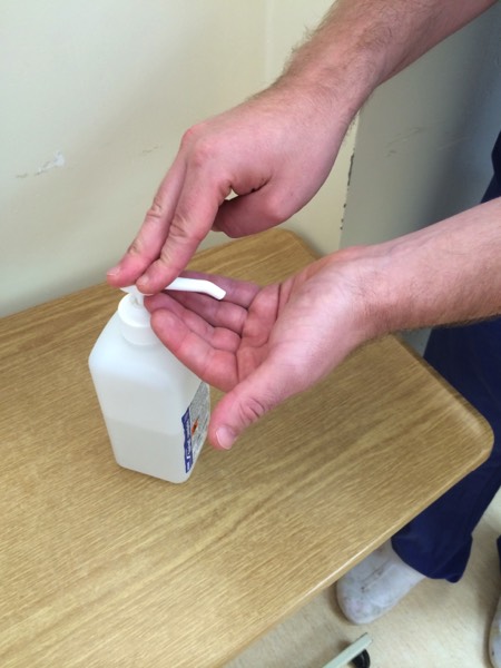
Wash your hands
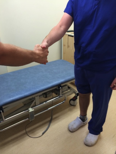
Introduce yourself to the patient
Explain that you would like to examine their knee joint
Check whether they are in any pain and which the ‘bad’ knee is
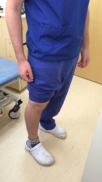
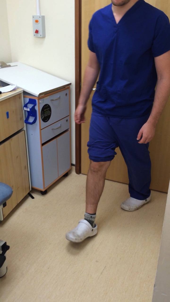
Look
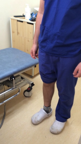
Inspect whilst standing
- Skin: Scars, swelling, erythema
- Muscle: Quadriceps wasting, effusions
- Bone & Joint: Valgus/varus deformity. Valgus =knees come together, varus = bow-legged
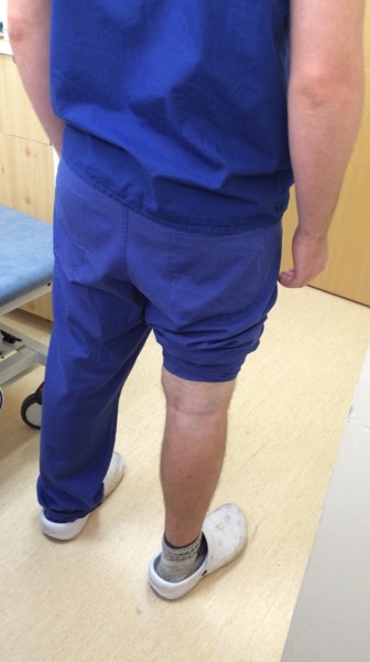
Check the front, back and side of their legs
Ask the patient to walk a short distance as a good screening test
You may wish to inspect the knee again with the patient lying down
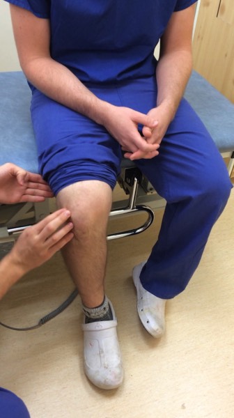
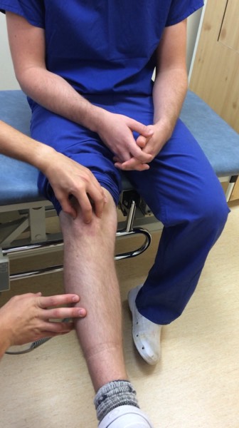
Feel
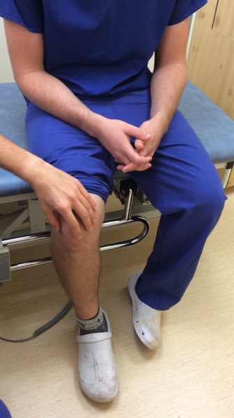
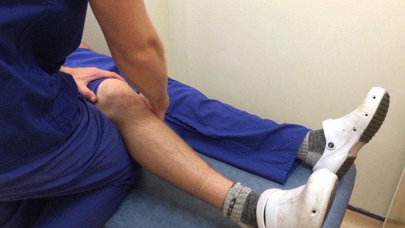
Sit the patient on the examination couch and look at their flexed knee
Palpate the joint line
With the patient sitting you may wish to test patella tracking
Place your thumb and middle finger either side of the patella and use your index finger to feel the top of the patella
Ask the patient to extend their knee from a flexed position
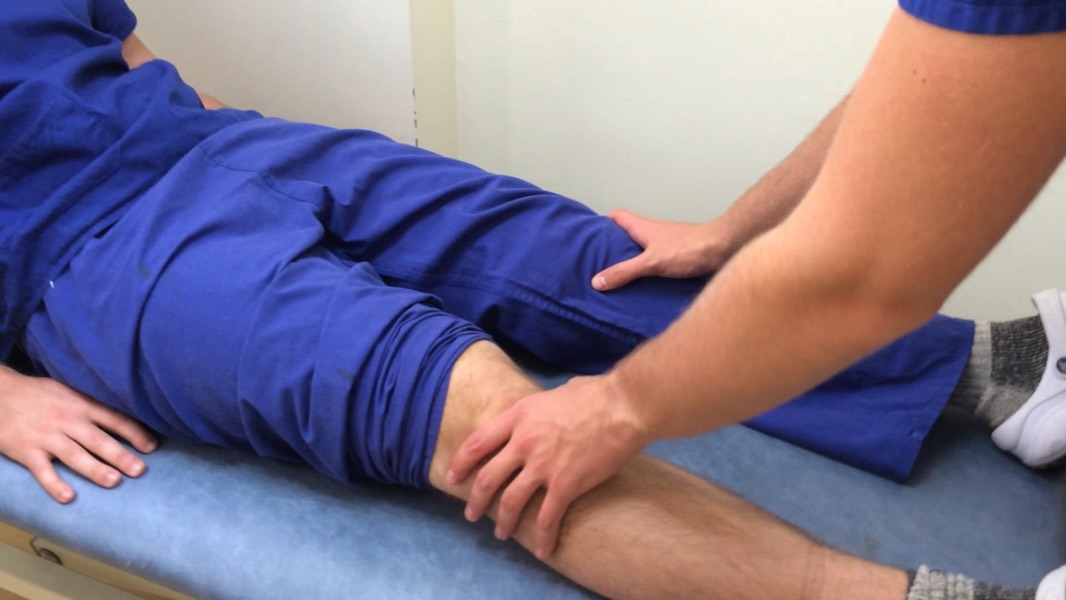
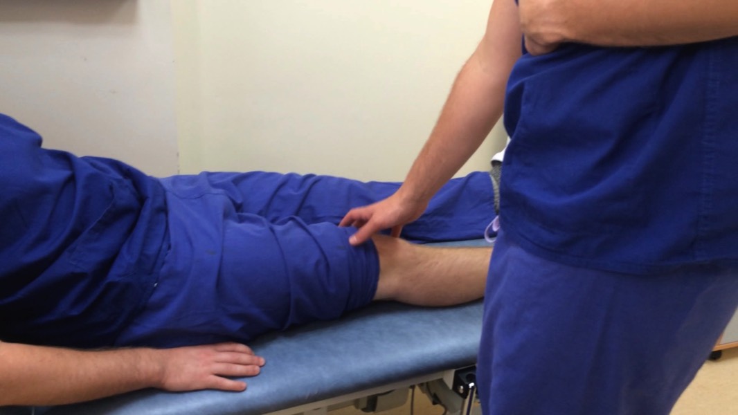
With the patient lying supine compare sides.
- Temperature: compare sides with the back of your hand
- Stroke test for effusions: empty the medial compartment with your hand by massaging fluid up and into the lateral side. Then apply pressure over lateral side whilst holding your other hand above the patella. Watch the medial gutter whilst doing this: it will balloon if there is an effusion.
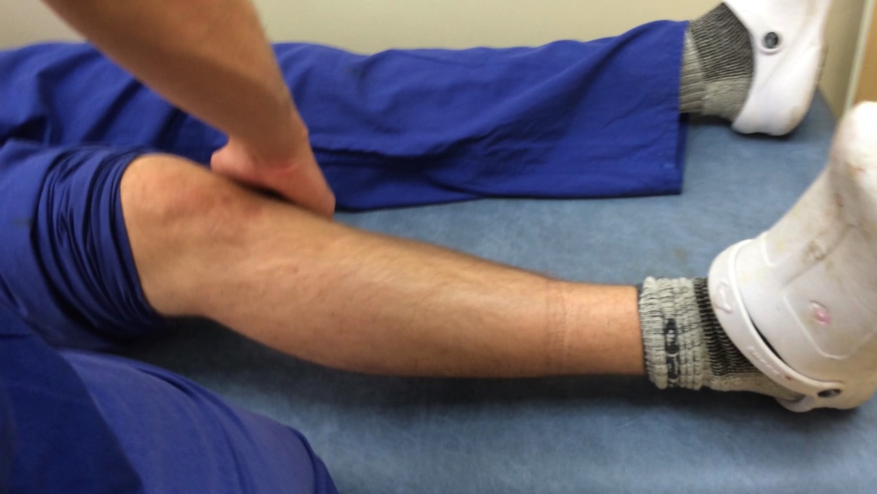
- Palpate Joint Margin: Bend knee to 90 degrees with foot on bed. Palpate joint line for tenderness (meniscal pathology), and the medial/lateral collaterals
- Examine popliteal fossa for cysts, aneurysm
- Move patella from side to side (knee slightly flexed)
Move
Active then passive movements. For passive movements keep one hand over the knee for crepitus.
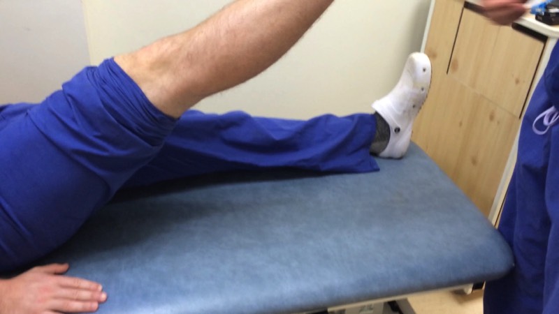
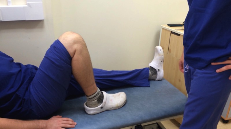
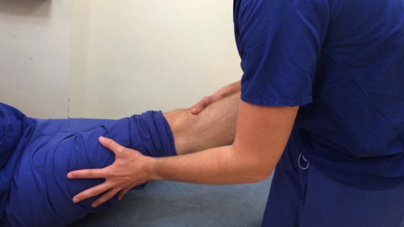
- Flexion (0-130°): bend their leg whilst putting a hand on their knee and feeling for crepitus. Their heel should be able to touch their buttocks
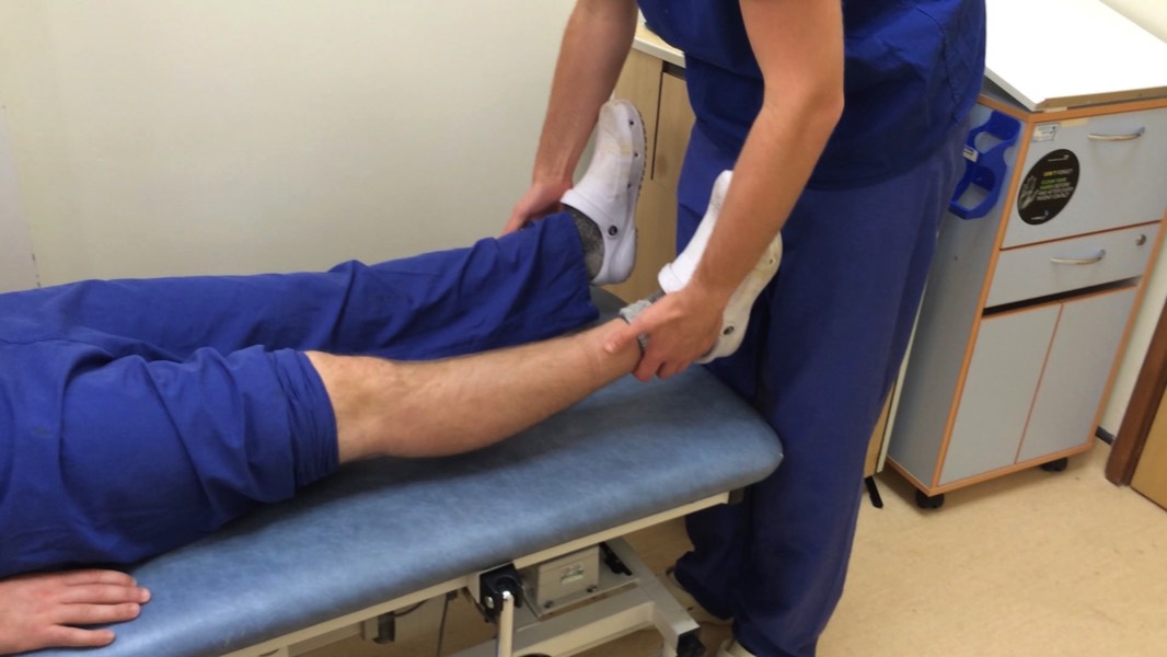
- Extension (0-15° ): Lift the patients leg in the air with your hand on their ankle (ask the patient to let you take their weight). If the leg extends fully then there is no fixed flexion
Special Tests
- Collateral ligaments: flex knee to 20 degrees and apply varus/valgus stress
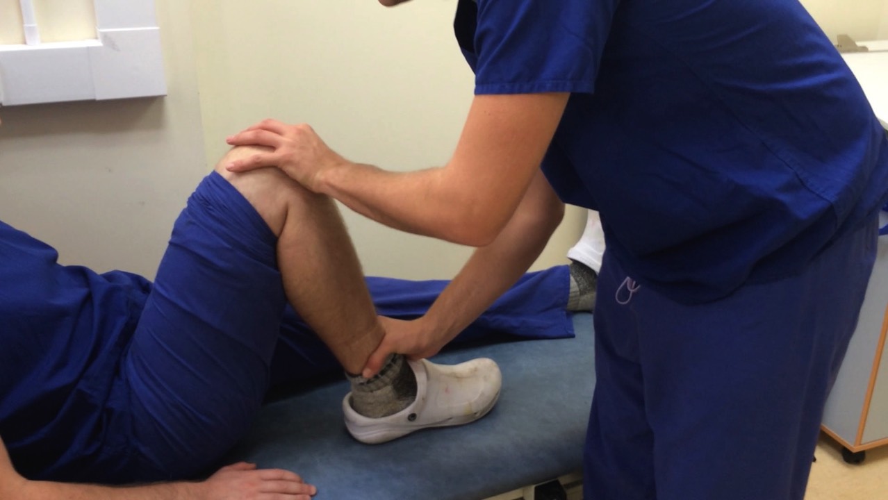
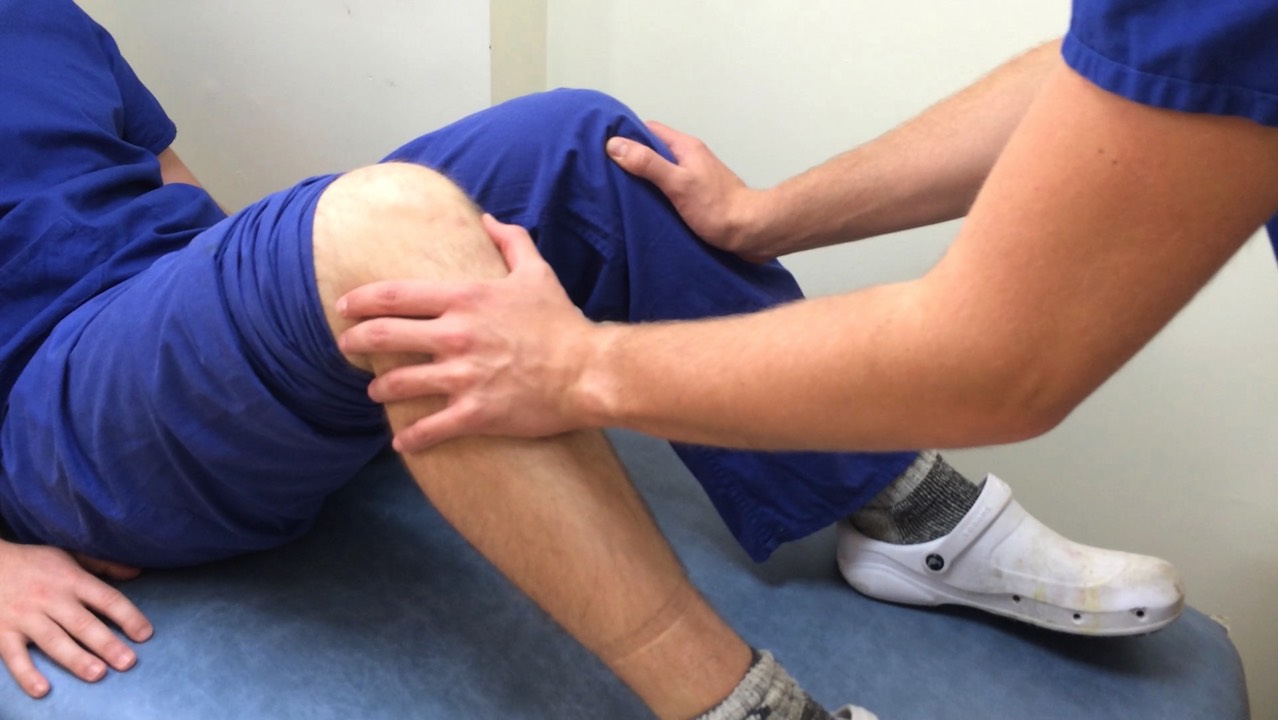
- Anterior/posterior draw test: With the knee at 90 degrees, look from the side for sag of tibia in relation to femur (tear of posterior cruciate). Now anchor the foot by sitting on the end of the toes (be very careful not to hurt the patient). Grab the leg below the knee with both hands and push it back (posterior draw for posterior cruciate tear) and pull forward (anterior draw test for anterior cruciate tear). The angle you should be drawing is parallel to the line of the femur. There should be slight but not excessive draw in each direction.
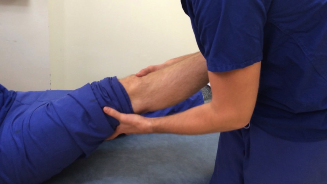
- Lachman's Test for anterior cruciate injury: bend the patient's leg to 30 degrees and grasp above the knee with one hand and below with the other. Apply slow pressure to the back of the proximal tibia (below the knee), increased laxness indicates injury.
- Pivot shift test for anterior cruciate rupture: Probably best to avoid in an exam as it can be painful. Flex the knee fully with the foot internally rotated. Apply slight valgus strain to the knee as it is slowly extended. A positive test is indicated by a click as the tibia shifts anteriorly on extension.
- McMurray's test for meniscal tears: McMurray's test is performed with the patient lying flat (non-weight bearing). Bend the knee and place one hand on the lateral side to stabilize the joint and provide a valgus stress. The other hand rotates the leg externally while extending the knee. A click is felt over the meniscus tear as the knee is brought from full flexion to 90 degrees of flexion.
Closing
Thank the patient
Wash your hands
State that you would like to:
Wash your hands
State that you would like to:
- Examine the joint above and below (hip and ankle)
- Get AP, lateral and skyline patella radiographs of the knee
- Assess the patient’s general fitness for surgery
P
Medical imagery licensed under Creative Commons Attribution-Share Alike license; sourced from Wikipedia
All other textual content, imagery, and website design copyright © 2014-22 MRCS Part B Questions all rights reserved.
Contact Us | Privacy Policy | Terms and Conditions